Bread mould (Penicillium sp.),SEM
Bildnummer 11525871
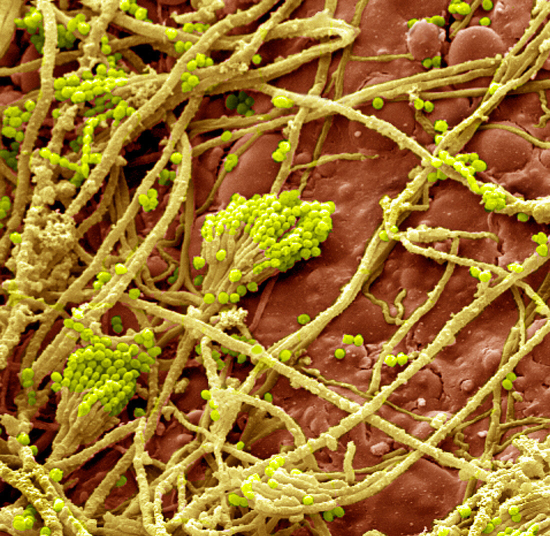
| Bread mould. Coloured scanning electron micrograph (SEM) of Penicillium sp. mould growing on bread. The yellow fibrous structures are hyphae,which make up the main body of the fungus. They penetrate the bread and absorb nutrients. The small green dots are the conidia,the spores of the mould. These are borne in clusters on specialised hyphae known as conidiophores. Magnification: x350 when printed 10cm wide | |
| Lizenzart: | Lizenzpflichtig |
| Credit: | Science Photo Library |
| Bildgröße: | 2552 px × 2487 px |
| Modell-Rechte: | nicht erforderlich |
| Eigentums-Rechte: | nicht erforderlich |
| Restrictions: | - |
Preise für dieses Bild ab 15 €
Universitäten & Organisationen
(Informationsmaterial Digital, Informationsmaterial Print, Lehrmaterial Digital etc.)
ab 15 €
Redaktionell
(Bücher, Bücher: Sach- und Fachliteratur, Digitale Medien (redaktionell) etc.)
ab 30 €
Werbung
(Anzeigen, Aussenwerbung, Digitale Medien, Fernsehwerbung, Karten, Werbemittel, Zeitschriften etc.)
ab 55 €
Handelsprodukte
(bedruckte Textilie, Kalender, Postkarte, Grußkarte, Verpackung etc.)
ab 75 €
Pauschalpreise
Rechtepakete für die unbeschränkte Bildnutzung in Print oder Online
ab 495 €