Helicobacter pylori bacteria
Bildnummer 11524033
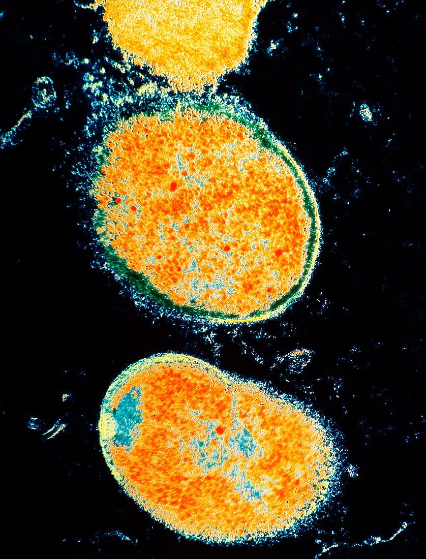
| Helicobacter pylori. Coloured transmission electron micrograph of two H. pylori bacteria in mucous of the human stomach. These bacteria were formerly called Campylobacter pyloridis. Here,the round cocci form of these Gram-negative bacteria is seen. They have flagellae for motility (not seen). The bacterial cell wall & cell contents are visible,surrounded by debris from epidermal cells of the stomach. Colonies of H. pylori occur on the stomach mucous membrane in people suffering gastritis,and this bacteria may be linked to stomach ulcers. H. pylori may also increase the risk of stomach tumours and gastric cancer. Magnification: x38,400 6x4. 5cm size | |
| Lizenzart: | Lizenzpflichtig |
| Credit: | Science Photo Library / Lounatmaa, Dr. Kari |
| Bildgröße: | 3691 px × 4843 px |
| Modell-Rechte: | nicht erforderlich |
| Eigentums-Rechte: | nicht erforderlich |
| Restrictions: | - |
Preise für dieses Bild ab 15 €
Universitäten & Organisationen
(Informationsmaterial Digital, Informationsmaterial Print, Lehrmaterial Digital etc.)
ab 15 €
Redaktionell
(Bücher, Bücher: Sach- und Fachliteratur, Digitale Medien (redaktionell) etc.)
ab 30 €
Werbung
(Anzeigen, Aussenwerbung, Digitale Medien, Fernsehwerbung, Karten, Werbemittel, Zeitschriften etc.)
ab 55 €
Handelsprodukte
(bedruckte Textilie, Kalender, Postkarte, Grußkarte, Verpackung etc.)
ab 75 €
Pauschalpreise
Rechtepakete für die unbeschränkte Bildnutzung in Print oder Online
ab 495 €