Model of the protein actin
Bildnummer 11521399
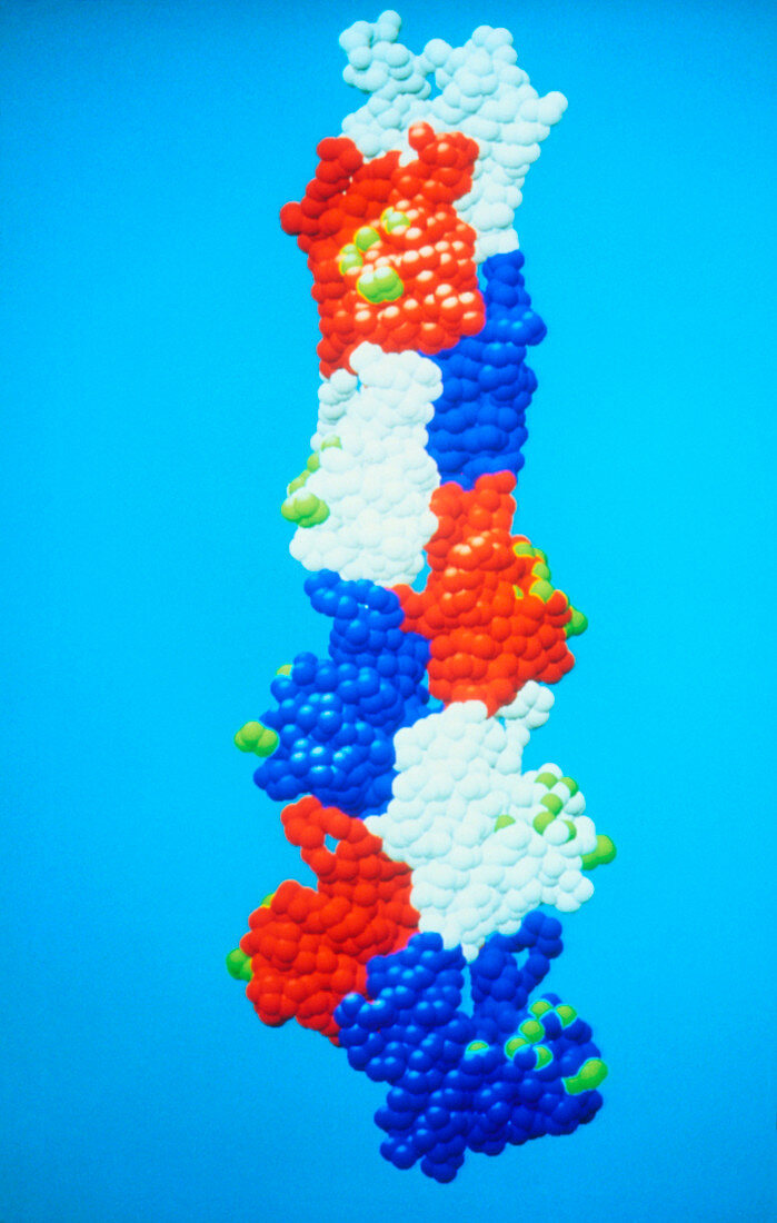
| Computer generated space-filling model of nine actin monomers from an F-actin helix,a polymer of the protein actin. For clarity each actin monomer (the repeated polymer unit) is shown in a different colour. Each amino-acid residue is represented by a sphere of radius 2.7 angstroms. In muscle cells,actin forms part of the thin filament,which cyclically interacts with the thick myosin filament to produce a mutual sliding that is the basis of muscle contraction. The green spheres are amino-acid residues that cross-link to the myosin in the actomyosin complex. The structure of the F-actin filament was determined using a technique called X-ray fibre diffraction | |
| Lizenzart: | Lizenzpflichtig |
| Credit: | Science Photo Library / Holmes, Dr. Kenneth |
| Bildgröße: | 2362 px × 3716 px |
| Modell-Rechte: | nicht erforderlich |
| Eigentums-Rechte: | nicht erforderlich |
| Restrictions: | - |
Preise für dieses Bild ab 15 €
Universitäten & Organisationen
(Informationsmaterial Digital, Informationsmaterial Print, Lehrmaterial Digital etc.)
ab 15 €
Redaktionell
(Bücher, Bücher: Sach- und Fachliteratur, Digitale Medien (redaktionell) etc.)
ab 30 €
Werbung
(Anzeigen, Aussenwerbung, Digitale Medien, Fernsehwerbung, Karten, Werbemittel, Zeitschriften etc.)
ab 55 €
Handelsprodukte
(bedruckte Textilie, Kalender, Postkarte, Grußkarte, Verpackung etc.)
ab 75 €
Pauschalpreise
Rechtepakete für die unbeschränkte Bildnutzung in Print oder Online
ab 495 €