Gold atoms on graphite substrate
Bildnummer 11518390
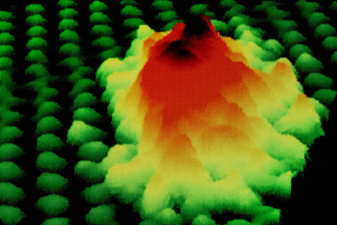
| Scanning tunnelling micrograph of gold atoms on a graphite substrate. The gold atoms,shown as yellow,red and brown,have formed an aggregate of 1x1. 5 nanometres at the base and about 3 atom layers thick. The graphite (carbon) atoms are shown as green. The scanning tunnelling microscope (STM) uses a fine point electrode,a few atoms thick at the tip. This is brought to within a few angstroms of the sample's surface. As the electron clouds of the tip and the sample atoms interact,a small 'tunnelling' current passes. The electrode scans across the surface,moving vertically to maintain the tunnelling current. This motion is then processed to produce a map such as this | |
| Lizenzart: | Lizenzpflichtig |
| Credit: | Science Photo Library / Plailly, Philippe |
| Bildgröße: | 4698 px × 3142 px |
| Modell-Rechte: | nicht erforderlich |
| Eigentums-Rechte: | nicht erforderlich |
| Restrictions: |
|
Preise für dieses Bild ab 15 €
Universitäten & Organisationen
(Informationsmaterial Digital, Informationsmaterial Print, Lehrmaterial Digital etc.)
ab 15 €
Redaktionell
(Bücher, Bücher: Sach- und Fachliteratur, Digitale Medien (redaktionell) etc.)
ab 30 €
Werbung
(Anzeigen, Aussenwerbung, Digitale Medien, Fernsehwerbung, Karten, Werbemittel, Zeitschriften etc.)
ab 55 €
Handelsprodukte
(bedruckte Textilie, Kalender, Postkarte, Grußkarte, Verpackung etc.)
ab 75 €
Pauschalpreise
Rechtepakete für die unbeschränkte Bildnutzung in Print oder Online
ab 495 €