16-cell human embryo on a pin
Bildnummer 11875070
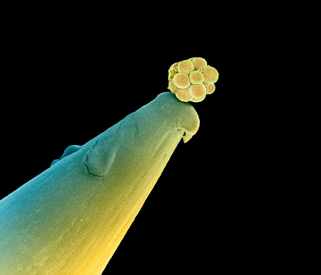
| Human embryo. Coloured scanning electron micrograph (SEM) of a human embryo at the 16-cell stage on the tip of a pin. The ball of cells (yellow) of the embryo is known as a morula,a cluster of almost identical,rounded cells,each containing a central nucleus. This 16-cell embryo is about three days old. It is at the early stage of transformation from a single cell to a human composed of millions of cells. The cells multiply by repeated cleavage divisions (mitosis) and will form a hollow ball of cells (the blastocyst). Development of the blastocyst occurs before the embryo implants into the wall of the uterus (womb). Magnification: x130 at 6x7cm size. Magnification: x450 at 8x10 inch size | |
| Lizenzart: | Lizenzpflichtig |
| Credit: | Science Photo Library / Nikas, Dr. Yorgos |
| Bildgröße: | 2800 px × 2400 px |
| Modell-Rechte: | nicht erforderlich |
| Eigentums-Rechte: | nicht erforderlich |
| Restrictions: | - |
Preise für dieses Bild ab 15 €
Universitäten & Organisationen
(Informationsmaterial Digital, Informationsmaterial Print, Lehrmaterial Digital etc.)
ab 15 €
Redaktionell
(Bücher, Bücher: Sach- und Fachliteratur, Digitale Medien (redaktionell) etc.)
ab 30 €
Werbung
(Anzeigen, Aussenwerbung, Digitale Medien, Fernsehwerbung, Karten, Werbemittel, Zeitschriften etc.)
ab 55 €
Handelsprodukte
(bedruckte Textilie, Kalender, Postkarte, Grußkarte, Verpackung etc.)
ab 75 €
Pauschalpreise
Rechtepakete für die unbeschränkte Bildnutzung in Print oder Online
ab 495 €
Keywords
- Bilder,
- Biologie,
- biologisch,
- Embryo,
- Embryologie,
- Entwicklung,
- entwicklungsgemäß,
- Fächer,
- farbig,
- gefärbt,
- gesund,
- Mensch,
- menschlicher Körper,
- mikroskopische Fotos,
- Morula,
- Nadel,
- Niemand,
- normal,
- Rasterelektronenmikroskop,
- rasterelektronenmikroskopische Aufnahme,
- REM,
- Reproduktion,
- schwarzer Hintergrund,
- Stammzellen,
- vergrößertes Bild,
- Zelle,
- Zellen