Bilder
Videos
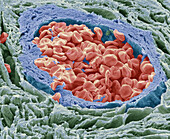
13674256 - Appendix artery, SEM
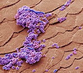
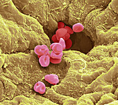
12489900 - Folliculitis bacteria, SEM
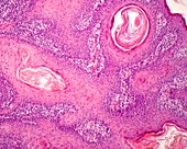
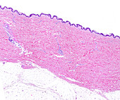
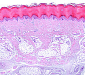
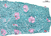
13387319 - Actinic keratosis, light micrograph

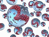
12659262 - Bacteria found on mobile phone, SEM

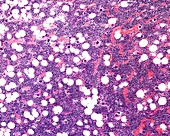
13523549 - Multiple myeloma, light micrograph
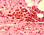
13736698 - Macrophages, light micrograph
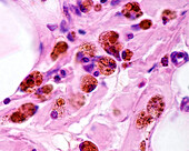
13736693 - Macrophages, light micrograph
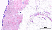
13673471 - Sole of the foot, light micrograph
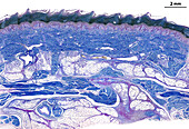
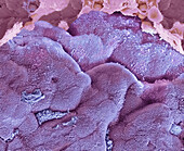
13673467 - Human skin, light micrograph
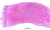
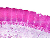
13673457 - Filiform papillae, light micrograph

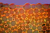
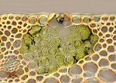
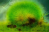
13634300 - Water milfoil, light micrograph
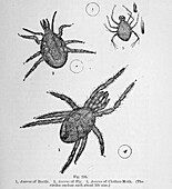
13634112 - Organisms under microscope, 19th century illustration
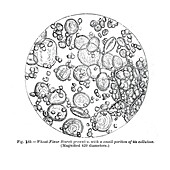
13634096 - Cells under microscope, 19th century illustration
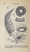
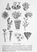
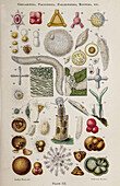
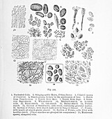
13634086 - Plant and fungi microscopy, 19th century illustration
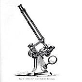
13634083 - Pillischer's larger Student's Microscope, illustration
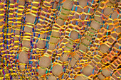
13633753 - Collenchyma in garden rhubarb stalk, light micrograph
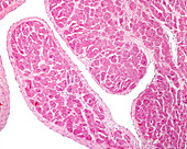
13613473 - Heart. Purkinje fibre, light micrograph
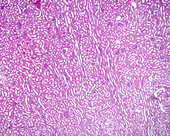
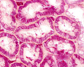
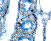
13586045 - Kidney cortex, light micrograph
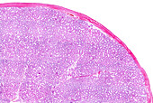
13673495 - Human testicle, light micrograph
13634117 - Organisms under microscope, 19th century illustration
13634090 - Cells under microscope, 19th century illustration
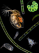
13620404 - Pond life, composite light micrograph

13613490 - Purkinje fibres in the heart, light micrograph
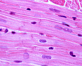
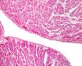
13613476 - Heart myocardium, light micrograph

13613474 - Purkinje fibres in the heart, light micrograph
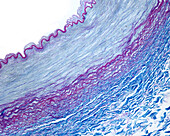
13613460 - Muscular artery, light micrograph
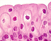
13586082 - Urinary bladder epithelium, light micrograph
13586076 - Kidney stained with Malloryâ??s trichrome, light micrograph
13586075 - Kidney cortex blood vessels, light micrograph
13586074 - Kidney medulla blood vessels, light micrograph

13586059 - Kidney stained with Polakâ??s silver method, light micrograph
13525553 - Yellow fever virus, TEM
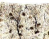
13243219 - Single cortical neuron, light micrograph
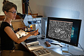
13732320 - Scientist identifying different species of moss
13732318 - Scientist identifying different species of moss
13686437 - Appendix, SEM
13673474 - Sole of the foot, light micrograph
13673468 - Human skin, light micrograph
13634109 - Organisms under microscope, 19th century illustration
13633941 - Field scabious stalk, light micrograph
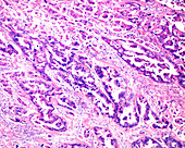
13620514 - Ovarian mucinous cystadenocarcinoma, light micrograph
13613453 - Human heart endocardium, light micrograph
13586069 - Kidney cortex, light micrograph
13586062 - Kidney development, light micrograph
13243195 - Cancer cell showing tubulin, light micrograph
14077786 - Fine Silk, light micrograph
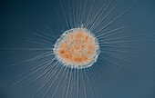
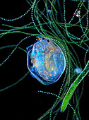
13674100 - Actinosphaerium heliozoan, light micrograph
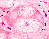
13673513 - Human fallopian tube, light micrograph
13673483 - Sole of the foot, light micrograph

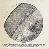
13673465 - Human nipple, light micrograph
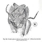
13634103 - Organisms under microscope, 19th century illustration
13634097 - Cells under microscope, 19th century illustration
13633186 - Blood vessel, SEM

13620511 - Mucosal crypts in human colon, light micrograph

13586053 - Kidney medulla, light micrograph
13585620 - Analysis of prehistoric flint tool
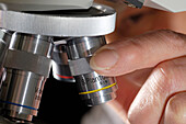
13524717 - Student adjusting microscope
13754802 - Sea slug eggs, light micrograph
13620513 - Ovarian mucinous cystadenocarcinoma, light micrograph
13954186 - Nitzschia sigmoidea and gomphonema algae, light micrograph
13620520 - Arterial thrombosis, light micrograph
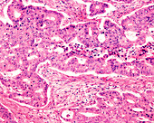
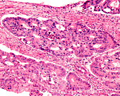
13620503 - Adenocarcinoma in human colon, light micrograph
13613454 - Human heart endocardium, light micrograph
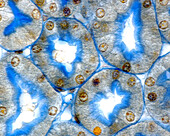
13586071 - Mitochondria in kidney convoluted tubules, light micrograph
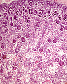
13426609 - Hematopoietic bone marrow, light micrograph
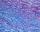
13673476 - Human skin dermis, light micrograph
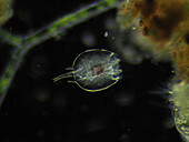
13634325 - Hydorus sp. water flea, light micrograph
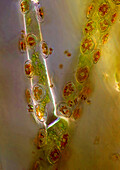
13634197 - Rye stoma, light micrograph

13634118 - Organisms under microscope, 19th century illustration
13634093 - Cells under microscope, 19th century illustration
13634089 - Cells under microscope, 19th century illustration
13620502 - Adenocarcinoma in human colon, light micrograph
13619930 - Chaetogaster diastrophus, light micrograph
13613462 - Muscular artery, light micrograph
13586077 - Kidney stained with Malloryâ??s trichrome, light micrograph
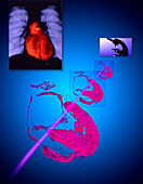
13585912 - Cardiology research, conceptual illustration
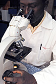
13523907 - Microscope use
13377892 - Messenger RNA vaccine, illustration
13634121 - Organisms under microscope, 19th century illustration
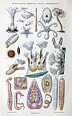
13634099 - Organisms under microscope, 19th century illustration
13619927 - Chaetogaster diastrophus, light micrograph

13613475 - Heart myocardium, light micrograph
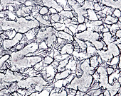
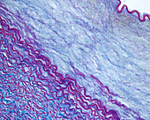
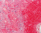
13601228 - Reticular fibres in human liver, light micrograph
13586083 - Urinary bladder epithelium, light micrograph
13586064 - Human foetal kidney corpuscle development, light micrograph
13525551 - Lassa virus budding, SEM
13673480 - Human nipple, light micrograph
13634315 - Green algae colony, light micrograph
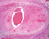
13620521 - Arterial thrombosis, light micrograph

13620504 - Adenocarcinoma in human colon, light micrograph
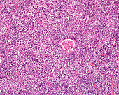
13620497 - Lymphosarcoma in human kidney, light micrograph
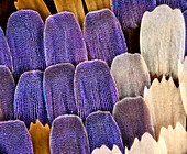
13620307 - Swallowtail butterfly wing scales, light micrograph
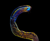
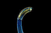
13619935 - Rotifer
13613461 - Muscular artery, light micrograph

13586065 - Kidney glomerulus basement membranes, light micrograph
13586063 - Human foetal kidney corpuscle development, light micrograph
13954184 - Cocconeis sp. algae, light micrograph
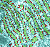
13755945 - Rough endoplasmic reticulum, TEM
13736699 - Lymph node macrophages, light micrograph
nächste Seite
Mikroskop Bilder ❘ Science Photo Library