Bilder
Videos

13736716 - Healthy eye and diabetic retinopathy, illustration
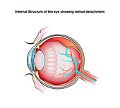
13736725 - Retinal detachment, illustration
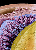
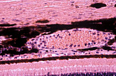
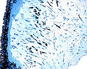
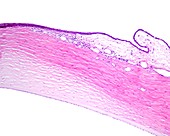
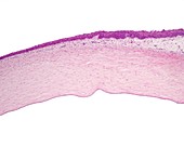
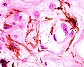
13756214 - Schlemm canal in eye, light micrograph

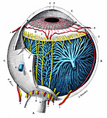
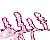
13732601 - Nerve structures of the retina, 1894 illustration

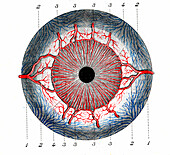
13686461 - Iris of the eye, SEM
13686439 - Iris of the eye, SEM

13736724 - Retinal detachment, illustration
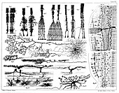
13732604 - Nerve structures of the retina, 1894 illustration
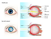
13736718 - Normal eye and eye with cataract, illustration
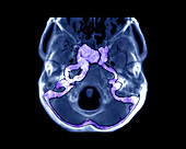
13599517 - Pulsatile exophthalmos, CT angiogram
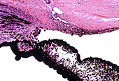
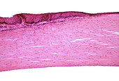
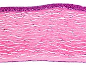
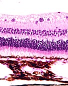
13756213 - Choroid and retina, light micrograph
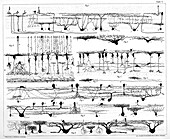
13732603 - Nerve structures of the retina, 1894 illustration
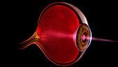
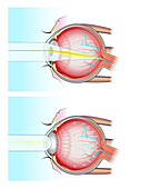
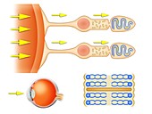
13585907 - Light entering human eye, illustration
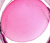
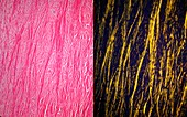
13756200 - Eye lens fibres, light micrograph
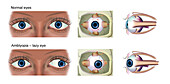
13736714 - Normal eye and amblyopia, illustration
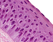
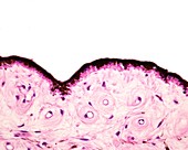
13756183 - Human cornea, light micrograph
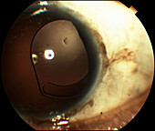
13613652 - Intraocular lens subluxation
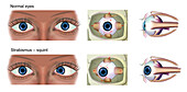
13736727 - Normal eye and strabismus, illustration
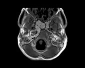
13599516 - Pulsatile exophthalmos, CT angiogram

13736715 - Healthy eye and diabetic retinopathy, illustration
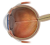
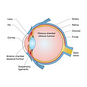
13733178 - Eye anatomy, illustration

13736135 - Eye blood vessels, illustration
13733177 - Eye anatomy, illustration
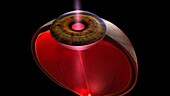
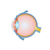
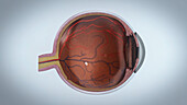
12640398 - Eyeball, cross section, illustration
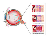
13736723 - Macular degeneration, illustration

13736722 - Macular degeneration, illustration
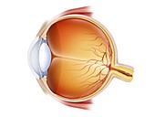
13585904 - Human eye anatomy, illustration
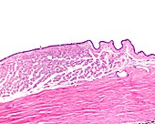
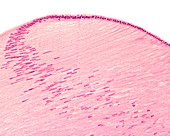
13426612 - Conjunctiva and sclera, light micrograph
13377636 - Eye anatomy, illustration

13756179 - Human cornea, light micrograph
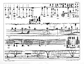
13732600 - Nerve structures of the retina, 1894 illustration
13416647 - Iris, light micrograph
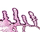
13756215 - Human ciliary body and iris, light micrograph
13426632 - Retina layers, light micrograph
13386588 - Human eyes, CT scan
13243917 - Ciliary body epithelium, light micrograph
13243908 - Iris stroma, light micrograph
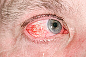
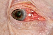
13951635 - Inflamed eye

13426627 - Iris sphincter muscle, light micrograph
13426623 - Limit between cornea and conjunctiva, light micrograph
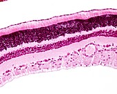
13243915 - Retina layers, light micrograph
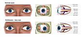
13736713 - Normal eye and amblyopia, illustration
13426624 - Ciliary body, light micrograph

13376764 - Eye anatomy, illustration
13243896 - Cornea layers, light micrograph
13243889 - Sclera, light micrograph
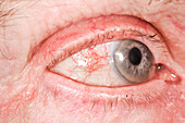
13951631 - Inflamed white of the eye
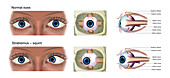
13736726 - Normal eye and strabismus, illustration
13736721 - Normal eye and eye with cataract, illustration
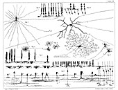
13732602 - Nerve structures of the retina, 1894 illustration
13386582 - Human eye, CT scan
13243905 - Cornea epithelium, light micrograph
13243900 - Choroid layer, light micrograph
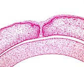
13243886 - Eye development, light micrograph
12987500 - Eye lens, light micrograph
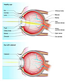
13736719 - Normal eye and eye with cataract, illustration
13756216 - Human conjunctiva and cornea, light micrograph

13736717 - Normal eye and eye with glaucoma, illustration
13243903 - Sclera, light micrograph
12378553 - Acute glaucoma in a blind eye

13243916 - Cat tapetum lucidum, light micrograph
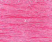
13243901 - Sclera, light micrograph

13736134 - Eye blood vessels, illustration

13732605 - Nerve structures of the retina, 1894 illustration
13377637 - Eye anatomy, illustration
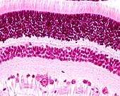
13243919 - Retina layers, light micrograph

13243897 - Cornea, light micrograph

12970993 - Eyeballs with headphones, illustration
12949097 - Cornea, light micrograph
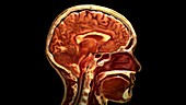
12647808 - Sagittal Section of Head
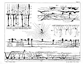
13732599 - Nerve structures of the retina, 1894 illustration
13632940 - Eye, illustration
13632938 - Eye, illustration
13377002 - Rear surface of iris, light micrograph

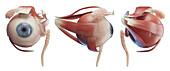
13376761 - Muscles of the eye, illustration
13243909 - Iris, light micrograph
13243907 - Back of iris, light micrograph
13243898 - Cornea and conjunctiva, light micrograph
12971338 - Biochemistry of the retina, illustration
12971292 - Eye anatomy, illustration
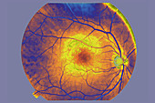
12636946 - Fundus of the Eye
13632939 - Eye, illustration
13599518 - Pulsatile exophthalmos, CT angiogram
13243914 - Lamina fusca, light micrograph
13223461 - Eyeballs, illustration
12949068 - Ciliary body, light micrograph
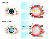
13736720 - Normal eye and eye with cataract, illustration
12949110 - Retina and choroid, light micrograph
13497122 - Cake pop shapes
13426625 - Ciliary processes, light micrograph
12949083 - Iris, light micrograph
12949072 - Melanocytes in iris stroma, light micrograph
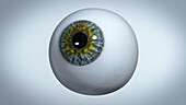
13962362 - Eyeball, illustration
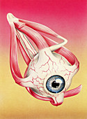
13376759 - Muscles of the eye, illustration
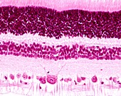
13243918 - Retina layers, light micrograph
12949098 - Embryonic fused eyelids, light micrograph
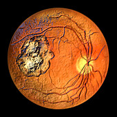
13956085 - Retinal scar caused by toxoplasmosis, illustration
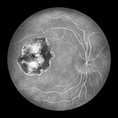
13956074 - Retinal scar caused by toxoplasmosis, illustration
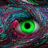
13674971 - Eye, abstract illustration
nächste Seite
Augapfel Bilder ❘ Science Photo Library