Spinal cord posterior horn, light micrograph
Bildnummer 12987521
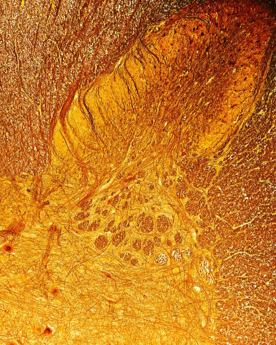
| Spinal cord posterior horn, light micrograph. Posterior or dorsal horn of spinal cord showing the substantia gelatinosa of Rolando (lamina II) covering the nucleus propius (lamina III). In the base of dorsal horn, the reticular formation (a mixture of grey and white matter, exclusive to the cervical spinal cord) can be seen. The substantia gelatinosa of Rolando is crossed by dorsal column-medial lemniscus tract fibres. Cajal's silver nitrate method. | |
| Lizenzart: | Lizenzpflichtig |
| Credit: | Science Photo Library / JOSE CALVO |
| Bildgröße: | 3072 px × 3840 px |
| Modell-Rechte: | nicht erforderlich |
| Eigentums-Rechte: | nicht erforderlich |
| Restrictions: | - |
Preise für dieses Bild ab 15 €
Universitäten & Organisationen
(Informationsmaterial Digital, Informationsmaterial Print, Lehrmaterial Digital etc.)
ab 15 €
Redaktionell
(Bücher, Bücher: Sach- und Fachliteratur, Digitale Medien (redaktionell) etc.)
ab 30 €
Werbung
(Anzeigen, Aussenwerbung, Digitale Medien, Fernsehwerbung, Karten, Werbemittel, Zeitschriften etc.)
ab 55 €
Handelsprodukte
(bedruckte Textilie, Kalender, Postkarte, Grußkarte, Verpackung etc.)
ab 75 €
Pauschalpreise
Rechtepakete für die unbeschränkte Bildnutzung in Print oder Online
ab 495 €
Keywords
- Biologie,
- biologisch,
- Gewebe,
- Histologie,
- histologisch,
- Lichtmikroskop,
- lichtmikroskopische Aufnahme,
- Mikrofotografie,
- Mikroskop,
- Mikroskopie,
- mikroskopisch,
- Nervensystem,
- Neuroimaging,
- Neurologie,
- neurologisch,
- Neuron,
- Neurowissenschaften,
- Niemand,
- Rückenmark,
- Silbernitrat,
- Soma,
- Zelle,
- zentrales Nervensystem