Intestinal epithelium, TEM
Bildnummer 12655586
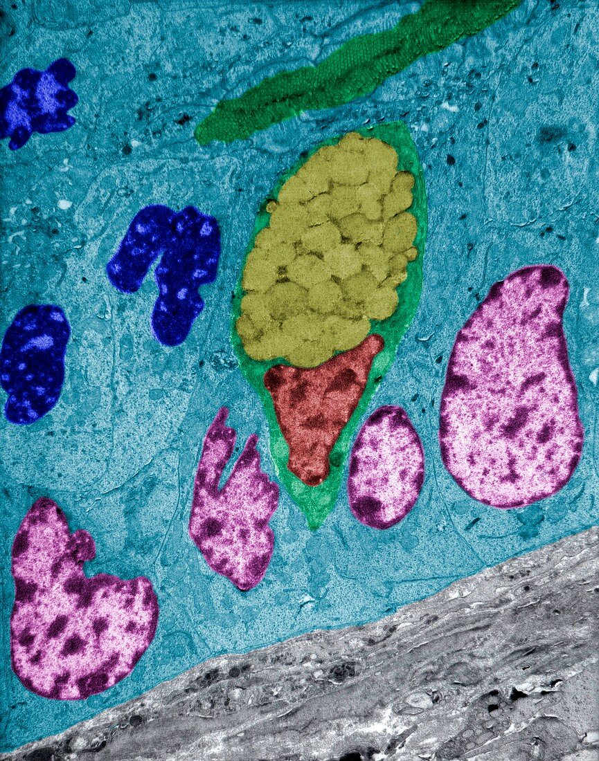
| False colour transmission electron micrograph (TEM) showing the intestinal epithelium of a Lieberkuhn crypt. The epithelium shows two cell types: enterocytes (cytoplasm-blue, nuclei-magenta, brush border-green) and goblet cells, which contain a nucleus (dark red), cytoplasm (green) and large mucous granules (dark yellow). Dark blue nuclei are in telophase, the final stage of mitosis. Mitotic cells are abundant at the bottom of the crypt. | |
| Lizenzart: | Lizenzpflichtig |
| Credit: | Science Photo Library / JOSE CALVO |
| Bildgröße: | 2731 px × 3472 px |
| Modell-Rechte: | nicht erforderlich |
| Eigentums-Rechte: | nicht erforderlich |
| Restrictions: | - |
Preise für dieses Bild ab 15 €
Universitäten & Organisationen
(Informationsmaterial Digital, Informationsmaterial Print, Lehrmaterial Digital etc.)
ab 15 €
Redaktionell
(Bücher, Bücher: Sach- und Fachliteratur, Digitale Medien (redaktionell) etc.)
ab 30 €
Werbung
(Anzeigen, Aussenwerbung, Digitale Medien, Fernsehwerbung, Karten, Werbemittel, Zeitschriften etc.)
ab 55 €
Handelsprodukte
(bedruckte Textilie, Kalender, Postkarte, Grußkarte, Verpackung etc.)
ab 75 €
Pauschalpreise
Rechtepakete für die unbeschränkte Bildnutzung in Print oder Online
ab 495 €