Brain areas affected by THC exposure, MRI scan
Bildnummer 13951498
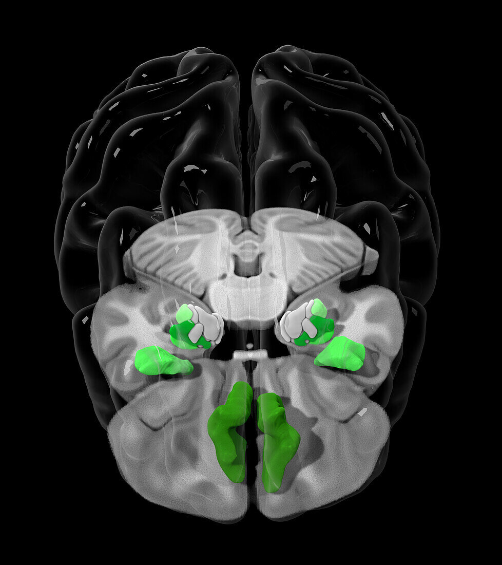
| Coloured magnetic resonance imaging (MRI) scan showing brain areas (green) that show differential microstructure organisation in people that are exposed to tetrahydrocannabinol (THC), the principal psychoactive constituent of cannabis. At bottom centre is the ventromedial prefrontal cortex (vmPFC), above that are the left and right agranular anterior insular cortexes and above them are the left and right amygdala. Changes in the microstructures of these areas have been shown to impact memory performance, negative intrusive thinking, and paternal substance abuse. | |
| Lizenzart: | Lizenzpflichtig |
| Credit: | Science Photo Library / MARK AND MARY STEVENS NEUROIMAGING AND INFORMATICS INSTITUTE |
| Bildgröße: | 3947 px × 4440 px |
| Modell-Rechte: | nicht erforderlich |
| Eigentums-Rechte: | nicht erforderlich |
| Restrictions: | - |
Preise für dieses Bild ab 15 €
Universitäten & Organisationen
(Informationsmaterial Digital, Informationsmaterial Print, Lehrmaterial Digital etc.)
ab 15 €
Redaktionell
(Bücher, Bücher: Sach- und Fachliteratur, Digitale Medien (redaktionell) etc.)
ab 30 €
Werbung
(Anzeigen, Aussenwerbung, Digitale Medien, Fernsehwerbung, Karten, Werbemittel, Zeitschriften etc.)
ab 55 €
Handelsprodukte
(bedruckte Textilie, Kalender, Postkarte, Grußkarte, Verpackung etc.)
ab 75 €
Pauschalpreise
Rechtepakete für die unbeschränkte Bildnutzung in Print oder Online
ab 495 €