White matter fibres, DTI MRI scan
Bildnummer 13950872
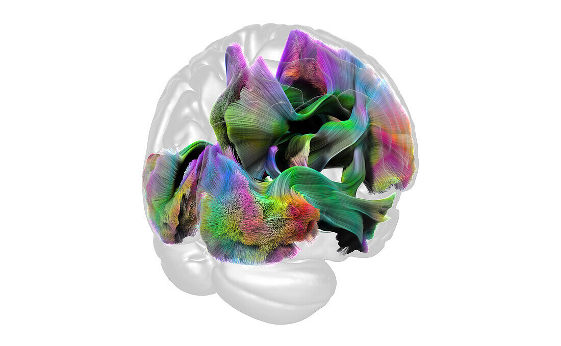
| Coloured diffusion tensor imaging (DTI) magnetic resonance imaging (MRI) scan showing white matter fibres in the brain. The back of the brain is at bottom left. The structures shown here are the fornix, inferior longitudinal fibres, cingulum hippocampal fibres, cingulum, inferior fronto-occipital fibres, thalamic prefrontal fibres and uncinate fasciculus pathway. Diffusion tensor imaging measures the direction of water diffusion, which in the brain reveals the orientation of nerve fibres. Red fibres have a left to right orientation, green a front to back orientation and blue an up and down orientation. | |
| Lizenzart: | Lizenzpflichtig |
| Credit: | Science Photo Library / MARK AND MARY STEVENS NEUROIMAGING AND INFORMATICS INSTITUTE |
| Bildgröße: | 5330 px × 3288 px |
| Modell-Rechte: | nicht erforderlich |
| Eigentums-Rechte: | nicht erforderlich |
| Restrictions: | - |
Preise für dieses Bild ab 15 €
Universitäten & Organisationen
(Informationsmaterial Digital, Informationsmaterial Print, Lehrmaterial Digital etc.)
ab 15 €
Redaktionell
(Bücher, Bücher: Sach- und Fachliteratur, Digitale Medien (redaktionell) etc.)
ab 30 €
Werbung
(Anzeigen, Aussenwerbung, Digitale Medien, Fernsehwerbung, Karten, Werbemittel, Zeitschriften etc.)
ab 55 €
Handelsprodukte
(bedruckte Textilie, Kalender, Postkarte, Grußkarte, Verpackung etc.)
ab 75 €
Pauschalpreise
Rechtepakete für die unbeschränkte Bildnutzung in Print oder Online
ab 495 €
Keywords
- 3D,
- Ballaststoff,
- cgi,
- digital generiert,
- Dreidimensional,
- dti,
- dti Scan,
- farbig,
- Fasern,
- Fornix,
- Gehirn,
- gesund,
- Hinter-,
- Hirnscan,
- Limbisches System,
- Magnetresonanztomografie,
- menschlicher Körper,
- Modell-,
- MRI,
- Nerven,
- Nerventrakt,
- Neuroimaging,
- Niemand,
- normal,
- posterior,
- Weg,
- Wege,
- weiße Substanz,
- weißer Hintergrund,
- zentrales Nervensystem