Cytokinesis, light micrograph
Bildnummer 13755891
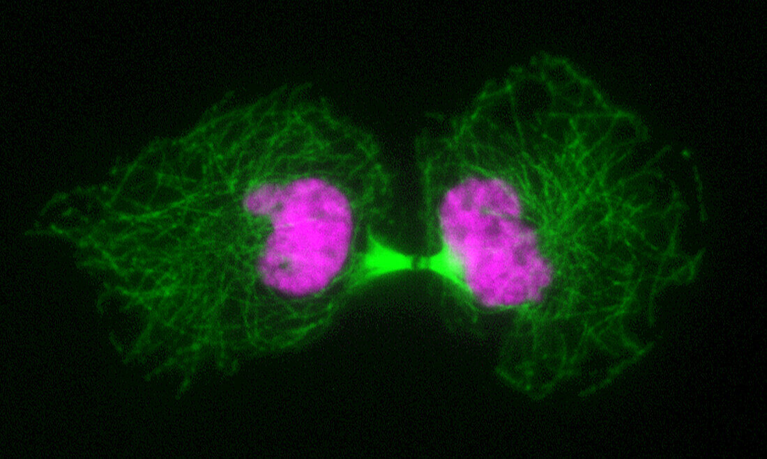
| Fluorescent light micrograph of a cell during cytokinesis (cell division). Fluorescent markers have been used to highlight alpha tubulin (green), a component of microtubules, and DNA (deoxyribonucleic acid, pink). At centre is a midbody, a bridge of cytoplasm between two separating cells. Division of a cell's cytoplasm follows shortly after division of the nucleus (mitosis). A contractile ring of actin filaments gradually narrows the bridge between the two cells until it breaks the microtubule fibres of the spindle (bright green) and divides the cells. The spindle is involved in division of chromosomes during mitosis, the formation of two daughter nuclei from one parent nucleus. | |
| Lizenzart: | Lizenzpflichtig |
| Credit: | Science Photo Library / DR. JUAN F. GIMENEZ-ABIAN |
| Bildgröße: | 3827 px × 2292 px |
| Modell-Rechte: | nicht erforderlich |
| Eigentums-Rechte: | nicht erforderlich |
| Restrictions: | - |
Preise für dieses Bild ab 15 €
Universitäten & Organisationen
(Informationsmaterial Digital, Informationsmaterial Print, Lehrmaterial Digital etc.)
ab 15 €
Redaktionell
(Bücher, Bücher: Sach- und Fachliteratur, Digitale Medien (redaktionell) etc.)
ab 30 €
Werbung
(Anzeigen, Aussenwerbung, Digitale Medien, Fernsehwerbung, Karten, Werbemittel, Zeitschriften etc.)
ab 55 €
Handelsprodukte
(bedruckte Textilie, Kalender, Postkarte, Grußkarte, Verpackung etc.)
ab 75 €
Pauschalpreise
Rechtepakete für die unbeschränkte Bildnutzung in Print oder Online
ab 495 €
Keywords
- alpha-Tubulin,
- Atomkern,
- Biologie,
- biologisch,
- Brücke,
- Desoxiribonukleinsäure,
- DNA,
- Fluoreszenz,
- fluoreszierend,
- Genetik,
- kopierend,
- lichtmikroskopische Aufnahme,
- Mikroskopie,
- Mikrotubuli,
- Mitose,
- mitotische Spindel,
- Niemand,
- Reproduktion,
- schwarzer Hintergrund,
- Separieren,
- Trennung,
- Zellbilogie,
- Zelle,
- Zytokinese,
- Zytologie,
- Zytologisch,
- Zytoplasma,
- Zytoskelett