Epidermis, light micrograph
Bildnummer 13732488
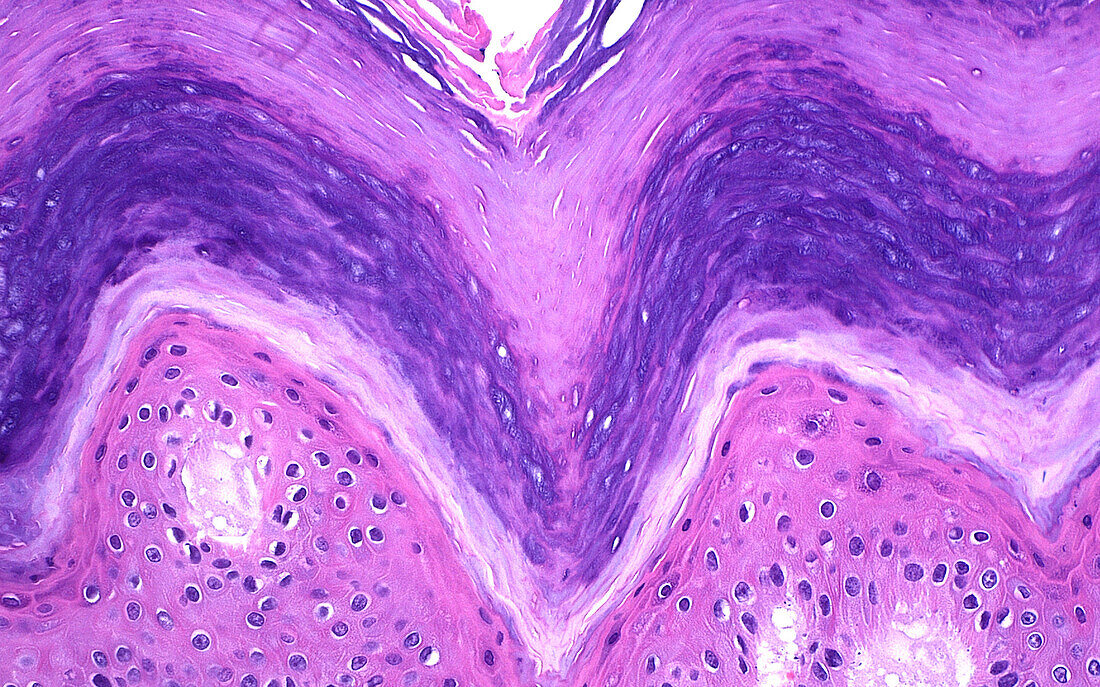
| Light micrograph of the epidermis (surface layer) of skin. The normal skin surface pokes up and down forming the so-called rete ridges, forming as the 'spiky' structure in seen in this picture. Layers of the epidermis include the spinous layer (lowermost) which has the maturing pink keratinocytes (skin cells), the corneal layer (uppermost) which is composed of acellular pink and purple keratin material, and the lucidum layer in between the spinous and corneal layers, which is the clear line seen in the picture. Haematoxylin and eosin stained tissue section. Magnification: 200x when printed at 10cm. | |
| Lizenzart: | Lizenzpflichtig |
| Credit: | Science Photo Library / ZIAD M. EL-ZAATARI |
| Bildgröße: | 5000 px × 3122 px |
| Modell-Rechte: | nicht erforderlich |
| Eigentums-Rechte: | nicht erforderlich |
| Restrictions: | - |
Preise für dieses Bild ab 15 €
Universitäten & Organisationen
(Informationsmaterial Digital, Informationsmaterial Print, Lehrmaterial Digital etc.)
ab 15 €
Redaktionell
(Bücher, Bücher: Sach- und Fachliteratur, Digitale Medien (redaktionell) etc.)
ab 30 €
Werbung
(Anzeigen, Aussenwerbung, Digitale Medien, Fernsehwerbung, Karten, Werbemittel, Zeitschriften etc.)
ab 55 €
Handelsprodukte
(bedruckte Textilie, Kalender, Postkarte, Grußkarte, Verpackung etc.)
ab 75 €
Pauschalpreise
Rechtepakete für die unbeschränkte Bildnutzung in Print oder Online
ab 495 €