Bladder in situ cancer, light micrograph
Bildnummer 13732385
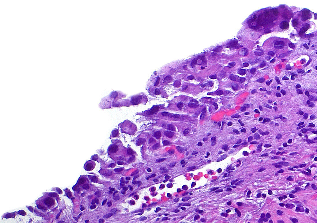
| Light micrograph of in situ urothelial carcinoma (CIS), which is a premalignant condition or one that has the potential to develop into invasive cancer. CIS is composed enlarged, hyperchromatic (dark staining), and irregular shaped cells. The picture shows cells of CIS spanning left bottom to right top of the image. The right upper to lower half of the image shows the benign connective tissue underneath the abnormal CIS cells. Haematoxylin and eosin stained tissue section. Magnification: 200x when printed at 10 cm. | |
| Lizenzart: | Lizenzpflichtig |
| Credit: | Science Photo Library / ZIAD M. EL-ZAATARI |
| Bildgröße: | 5000 px × 3517 px |
| Modell-Rechte: | nicht erforderlich |
| Eigentums-Rechte: | nicht erforderlich |
| Restrictions: | - |
Preise für dieses Bild ab 15 €
Universitäten & Organisationen
(Informationsmaterial Digital, Informationsmaterial Print, Lehrmaterial Digital etc.)
ab 15 €
Redaktionell
(Bücher, Bücher: Sach- und Fachliteratur, Digitale Medien (redaktionell) etc.)
ab 30 €
Werbung
(Anzeigen, Aussenwerbung, Digitale Medien, Fernsehwerbung, Karten, Werbemittel, Zeitschriften etc.)
ab 55 €
Handelsprodukte
(bedruckte Textilie, Kalender, Postkarte, Grußkarte, Verpackung etc.)
ab 75 €
Pauschalpreise
Rechtepakete für die unbeschränkte Bildnutzung in Print oder Online
ab 495 €