Venule, light micrograph
Bildnummer 13686894
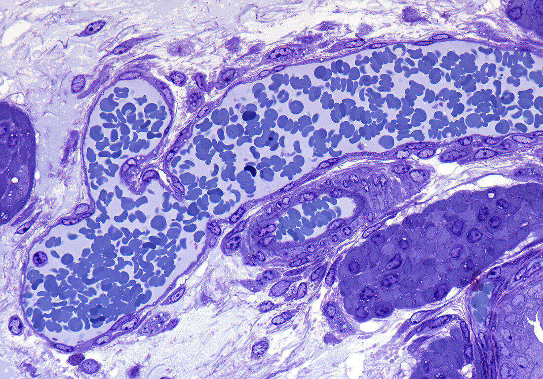
| Venule, light micrograph. Longitudinal section through a venule with a smaller joining tributary vessel. Venules have a thin wall of inner endothelium external to which are fibroblasts and mesenchymal cells collectively referred to as pericytes. An adjacent arteriole of smaller calibre shows a thicker wall comprised chiefly of smooth muscle. Epoxy resin section, Toluidine blue stain. Magnification: x350 when width printed at 10cm. | |
| Lizenzart: | Lizenzpflichtig |
| Credit: | Science Photo Library / Microscape |
| Bildgröße: | 4961 px × 3466 px |
| Modell-Rechte: | nicht erforderlich |
| Eigentums-Rechte: | nicht erforderlich |
| Restrictions: | - |
Preise für dieses Bild ab 15 €
Universitäten & Organisationen
(Informationsmaterial Digital, Informationsmaterial Print, Lehrmaterial Digital etc.)
ab 15 €
Redaktionell
(Bücher, Bücher: Sach- und Fachliteratur, Digitale Medien (redaktionell) etc.)
ab 30 €
Werbung
(Anzeigen, Aussenwerbung, Digitale Medien, Fernsehwerbung, Karten, Werbemittel, Zeitschriften etc.)
ab 55 €
Handelsprodukte
(bedruckte Textilie, Kalender, Postkarte, Grußkarte, Verpackung etc.)
ab 75 €
Pauschalpreise
Rechtepakete für die unbeschränkte Bildnutzung in Print oder Online
ab 495 €