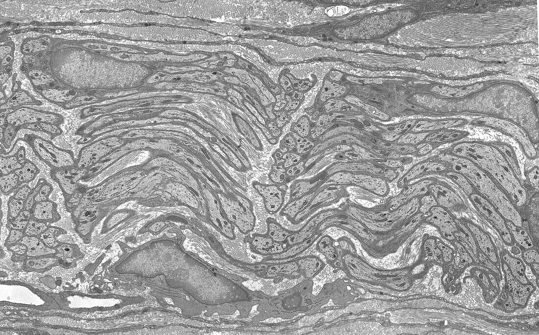
Nerve, TEM
Bildnummer 13620790

| Transmission electron micrograph (TEM) of the ultrastructure of an unmyelinated nerve showing multiple axons mostly sectioned longitudinally. The bundle of axons is supported (above and below) by connective tissues. A nucleus of a connective tissue fibroblast is seen at lower left. Each axon is invested by thin cytoplasm of Schwann cells with two nuclei of these cells seen at upper left and upper right. Collagen fibres are noted between the axons and represent the endoneurium. Magnification: x2, 800 when width printed at 10cm. | |
| Lizenzart: | Lizenzpflichtig |
| Credit: | Science Photo Library / Microscape |
| Bildgröße: | 5315 px × 3311 px |
| Modell-Rechte: | nicht erforderlich |
| Eigentums-Rechte: | nicht erforderlich |
| Restrictions: | - |
Preise für dieses Bild ab 15 €
Universitäten & Organisationen
(Informationsmaterial Digital, Informationsmaterial Print, Lehrmaterial Digital etc.)
ab 15 €
Redaktionell
(Bücher, Bücher: Sach- und Fachliteratur, Digitale Medien (redaktionell) etc.)
ab 30 €
Werbung
(Anzeigen, Aussenwerbung, Digitale Medien, Fernsehwerbung, Karten, Werbemittel, Zeitschriften etc.)
ab 55 €
Handelsprodukte
(bedruckte Textilie, Kalender, Postkarte, Grußkarte, Verpackung etc.)
ab 75 €
Pauschalpreise
Rechtepakete für die unbeschränkte Bildnutzung in Print oder Online
ab 495 €
