Microvilli, TEM
Bildnummer 13620785
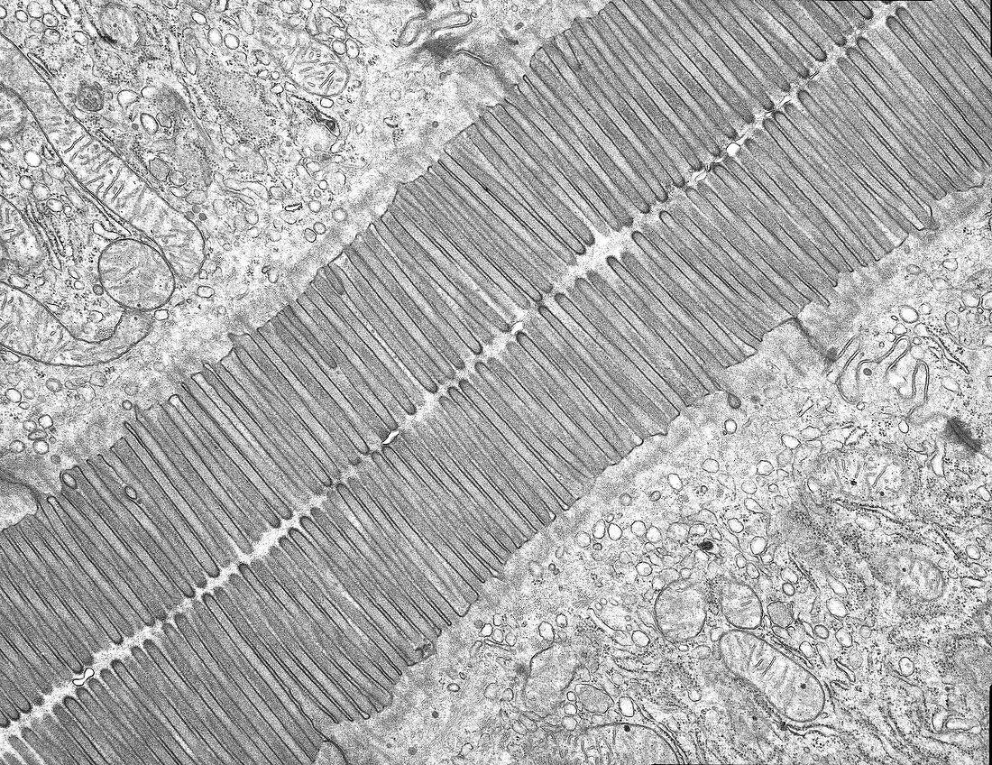
| Transmission electron micrograph (TEM) of the ultrastructure of apposed microvilli of two enterocytes of the small intestine epithelium. Microvilli are finger-like projections of the apical surface membrane of intestinal absorptive cells. They are about 1 micrometre in length with a core of actin filaments and greatly increase the membrane surface area for absorption of nutrients in the intestinal lumen. Fluid secretion by enterocytes also occurs across the microvilli. Magnification: x10, 000 when width printed at 10cm. | |
| Lizenzart: | Lizenzpflichtig |
| Credit: | Science Photo Library / Microscape |
| Bildgröße: | 5079 px × 3916 px |
| Modell-Rechte: | nicht erforderlich |
| Eigentums-Rechte: | nicht erforderlich |
| Restrictions: | - |
Preise für dieses Bild ab 15 €
Universitäten & Organisationen
(Informationsmaterial Digital, Informationsmaterial Print, Lehrmaterial Digital etc.)
ab 15 €
Redaktionell
(Bücher, Bücher: Sach- und Fachliteratur, Digitale Medien (redaktionell) etc.)
ab 30 €
Werbung
(Anzeigen, Aussenwerbung, Digitale Medien, Fernsehwerbung, Karten, Werbemittel, Zeitschriften etc.)
ab 55 €
Handelsprodukte
(bedruckte Textilie, Kalender, Postkarte, Grußkarte, Verpackung etc.)
ab 75 €
Pauschalpreise
Rechtepakete für die unbeschränkte Bildnutzung in Print oder Online
ab 495 €