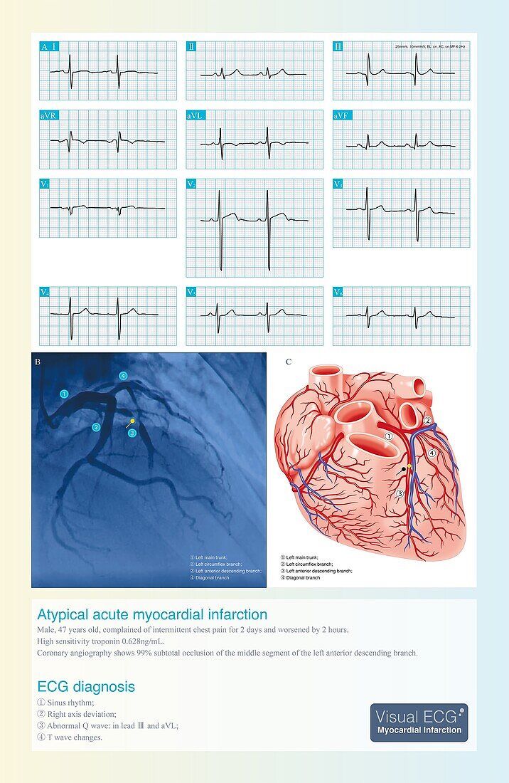
Acute coronary syndrome, illustration
Bildnummer 13453431

| Electrocardiogram (ECG) illustration from a 47 year old man admitted to hospital when he did not pay attention to intermittent chest pain for 2 days. High-sensitivity troponin is positive. Coronary angiography revealed 99% subtotal occlusion of the middle segment of left anterior descending branch. The coronary artery lesions were mainly located in the middle of left anterior descending artery. The left ventricular free wall myocardium is supplied by the developed diagonal branch and left circumflex branch, which provides ischemic protection, so the ECG myocardial infarction pattern is not typical. | |
| Lizenzart: | Lizenzpflichtig |
| Credit: | Science Photo Library / CHONGQING TUMI TECHNOLOGY LTD |
| Bildgröße: | 2800 px × 4302 px |
| Modell-Rechte: | nicht erforderlich |
| Eigentums-Rechte: | nicht erforderlich |
| Restrictions: | - |
Preise für dieses Bild ab 15 €
Universitäten & Organisationen
(Informationsmaterial Digital, Informationsmaterial Print, Lehrmaterial Digital etc.)
ab 15 €
Redaktionell
(Bücher, Bücher: Sach- und Fachliteratur, Digitale Medien (redaktionell) etc.)
ab 30 €
Werbung
(Anzeigen, Aussenwerbung, Digitale Medien, Fernsehwerbung, Karten, Werbemittel, Zeitschriften etc.)
ab 55 €
Handelsprodukte
(bedruckte Textilie, Kalender, Postkarte, Grußkarte, Verpackung etc.)
ab 75 €
Pauschalpreise
Rechtepakete für die unbeschränkte Bildnutzung in Print oder Online
ab 495 €
