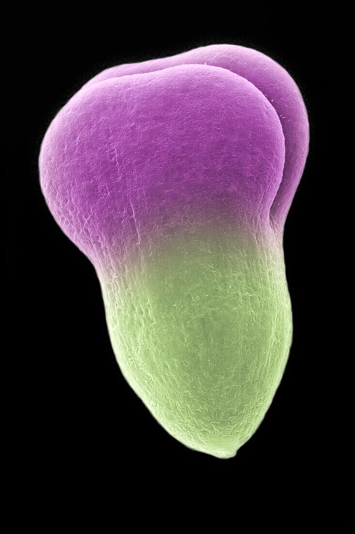
Developing embryo of Brassica campestris
Bildnummer 13435275

| Scanning electron micrograph of a developing embryo of the turnip, Brassica campestris. The picture shows a 1mm long embryo that was dissected from an immature seed. The upper regions (mauve) are the developing cotyledons (seed leaves); the lower region (pale green) will grow to form the root. The shoot meristem of the new plant is unseen, between the two cotyledons. This highly polarised structure developed over a period of less than three weeks from the fertilisation of one egg cell within the ovary of a flower. An embryo of this size has much further to grow, fed by the contents of the embryo sac. At maturity it becomes encased within a hard coat, as the contents of a dry viable seed. With favourable conditions of moisture and warmth, fed by food stores in the cotyledons, the shoot, root and internal vascular system begin to grow, breaking out of the seed coat to produce a new plant. | |
| Lizenzart: | Lizenzpflichtig |
| Credit: | Science Photo Library / Burgess, Dr. Jeremy |
| Bildgröße: | 3468 px × 5212 px |
| Modell-Rechte: | nicht erforderlich |
| Restrictions: | - |
Preise für dieses Bild ab 15 €
Universitäten & Organisationen
(Informationsmaterial Digital, Informationsmaterial Print, Lehrmaterial Digital etc.)
ab 15 €
Redaktionell
(Bücher, Bücher: Sach- und Fachliteratur, Digitale Medien (redaktionell) etc.)
ab 30 €
Werbung
(Anzeigen, Aussenwerbung, Digitale Medien, Fernsehwerbung, Karten, Werbemittel, Zeitschriften etc.)
ab 55 €
Handelsprodukte
(bedruckte Textilie, Kalender, Postkarte, Grußkarte, Verpackung etc.)
ab 75 €
Pauschalpreise
Rechtepakete für die unbeschränkte Bildnutzung in Print oder Online
ab 495 €
