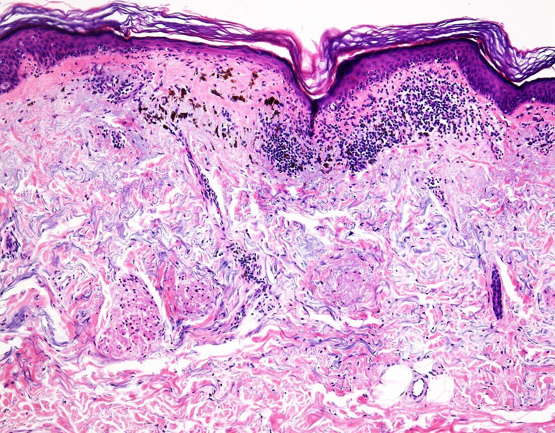
Melanoma with complete regression, light micrograph
Bildnummer 13403735

| Melanoma with complete regression, light micrograph. Histologic evidence of partial regression is seen in 10-35% of primary cutaneous melanomas. In a small number of cases, melanomas regress completely after giving rise to nodal or distant metastases. In 5-15% of cases of metastatic melanoma, the primary tumour is never found, presumably due to complete regression. Clinically, regression appears as macular gray, white, or pink areas with granularity. The regressed areas show a dermal lymphocytic infiltrate, absence of atypical melanocytic nests from the epidermis overlying the dermal infiltrate, epidermal atrophy with flattening of rete ridges, and pigment-laden macrophages. All of these features are seen in this image. | |
| Lizenzart: | Lizenzpflichtig |
| Credit: | Science Photo Library / WEBPATHOLOGY |
| Bildgröße: | 4096 px × 3200 px |
| Modell-Rechte: | nicht erforderlich |
| Eigentums-Rechte: | nicht erforderlich |
| Restrictions: | - |
Preise für dieses Bild ab 15 €
Universitäten & Organisationen
(Informationsmaterial Digital, Informationsmaterial Print, Lehrmaterial Digital etc.)
ab 15 €
Redaktionell
(Bücher, Bücher: Sach- und Fachliteratur, Digitale Medien (redaktionell) etc.)
ab 30 €
Werbung
(Anzeigen, Aussenwerbung, Digitale Medien, Fernsehwerbung, Karten, Werbemittel, Zeitschriften etc.)
ab 55 €
Handelsprodukte
(bedruckte Textilie, Kalender, Postkarte, Grußkarte, Verpackung etc.)
ab 75 €
Pauschalpreise
Rechtepakete für die unbeschränkte Bildnutzung in Print oder Online
ab 495 €
Keywords
- Dermatologie,
- dermatologisch,
- Epidermis,
- Hautkrebs,
- Histopathologie,
- histopathologisch,
- Krebs,
- krebsartig,
- Lichtmikroskop,
- lichtmikroskopische Aufnahme,
- maligne,
- malignes Melanom,
- Malignom,
- Melanom,
- Melanozyten,
- Metastase,
- Metastasen,
- Neoplasma,
- Onkologie,
- Pathologie,
- pathologisch,
- Sonnenbrand,
- sonnengeschädigt,
- Tumor
