Craniopharyngioma, light micrograph
Bildnummer 13296449
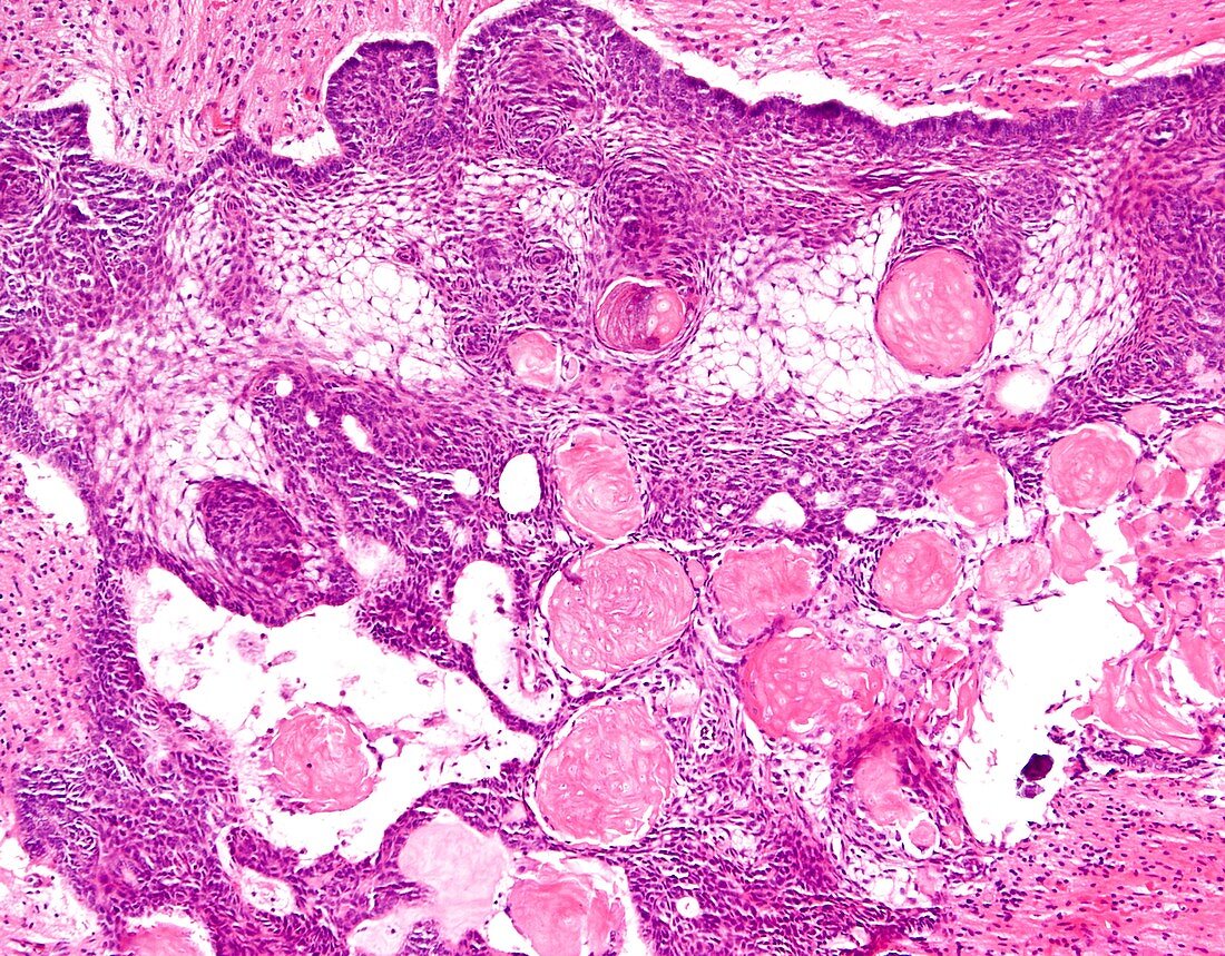
| Craniopharyngioma, light micrograph. Craniopharyngioma is a WHO Grade 1 epithelial tumour usually seen in the sellar/suprasellar region. It arises most likely from the remnants of Rathke's pouch. Some cases may have their origin in misplaced odontogenic rests along pituitary stalk. Grossly, they are often solid and cystic with areas of calcification. Craniopharyngiomas are subdivided into two types based on morphologic features: 1) Adamantinomatous (90% of cases) seen mostly in children and 2) Papillary (10% of cases) seen mostly in adults. The image shows adamantinomatous type with most of the key diagnostic features: a) sheets of squamous epithelial cells with peripheral palisading (which is best seen along the top of the image); b) a loose meshwork of epithelial cells called the stellate reticulum; c) nodules of anucleated squames (ghost cells) with brightly eosinophilic cytoplasm termed wet keratin. Wet keratin is considered diagnostic even in the absence of viable epithelium. | |
| Lizenzart: | Lizenzpflichtig |
| Credit: | Science Photo Library / WEBPATHOLOGY |
| Bildgröße: | 4096 px × 3200 px |
| Modell-Rechte: | nicht erforderlich |
| Eigentums-Rechte: | nicht erforderlich |
| Restrictions: | - |
Preise für dieses Bild ab 15 €
Universitäten & Organisationen
(Informationsmaterial Digital, Informationsmaterial Print, Lehrmaterial Digital etc.)
ab 15 €
Redaktionell
(Bücher, Bücher: Sach- und Fachliteratur, Digitale Medien (redaktionell) etc.)
ab 30 €
Werbung
(Anzeigen, Aussenwerbung, Digitale Medien, Fernsehwerbung, Karten, Werbemittel, Zeitschriften etc.)
ab 55 €
Handelsprodukte
(bedruckte Textilie, Kalender, Postkarte, Grußkarte, Verpackung etc.)
ab 75 €
Pauschalpreise
Rechtepakete für die unbeschränkte Bildnutzung in Print oder Online
ab 495 €
Keywords
- abnormal,
- Adamantinomatous,
- Cholesterol Kristalle,
- ctnnb1,
- Epilepsie,
- Gehirn,
- Histologie,
- histologisch,
- Histopathologie,
- histopathologisch,
- Karzinom,
- Kondition,
- Krankheit,
- Krebs,
- krebsartig,
- Lichtmikroskop,
- lichtmikroskopische Aufnahme,
- maligne,
- Malignom,
- Neurologie,
- neurologisch,
- Niemand,
- Onkologie,
- pädiatrisch,
- Pathologie,
- pathologisch,
- Störung,
- Tumor,
- ungesund,
- zentrales Nervensystem