Mitotic cell division, SEM-TEM comparison
Bildnummer 12948546
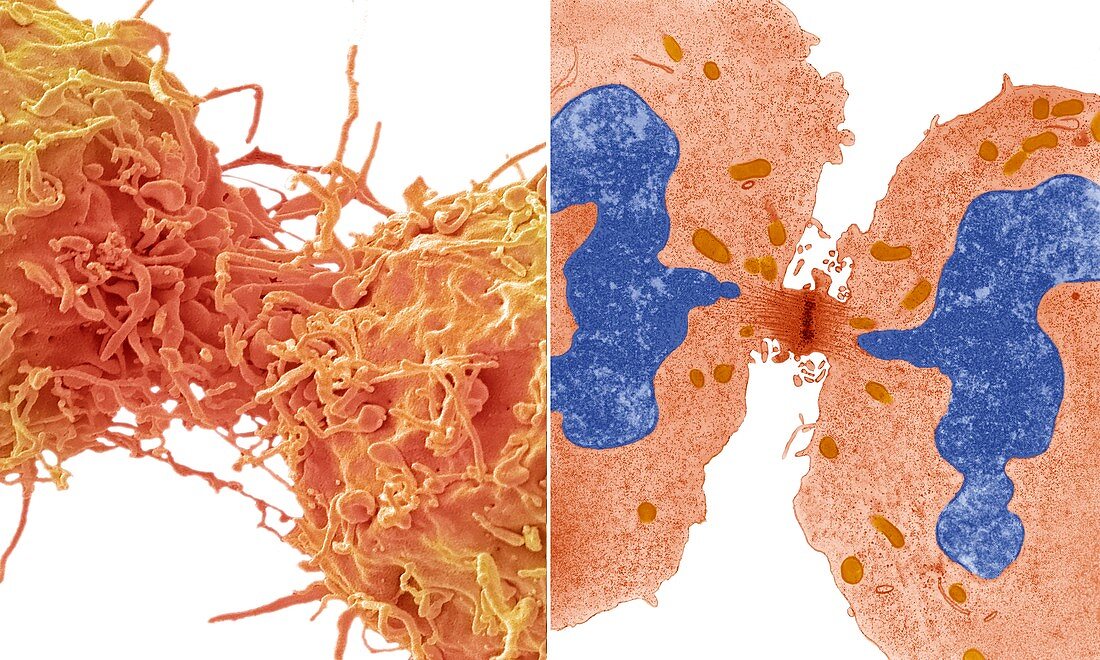
| Mitotic cell division. Comparison between a scanning electron micrograph (SEM, left) and transmission electron micrograph (TEM, right) of a cancer cell dividing by mitosis, the asexual replication of a cell into two new daughter cells. This is late telophase, the fourth stage of mitotic division, when the cell's genetic material (dark blue in TEM) has been separated via the spindle (centre) into two identical populations. The nuclear membranes have reformed around the genetic material (TEM) and the process of cytokinesis, the separation of the two new daughter cells, will shortly occur. Magnification: x10000 at 10 centimetres wide. For a series of comparisons between SEMs and TEMs see images C047/7006 to C047/7034. | |
| Lizenzart: | Lizenzpflichtig |
| Credit: | Science Photo Library / Gschmeissner, Steve |
| Bildgröße: | 7620 px × 4572 px |
| Modell-Rechte: | nicht erforderlich |
| Eigentums-Rechte: | nicht erforderlich |
| Restrictions: | - |
Preise für dieses Bild ab 15 €
Universitäten & Organisationen
(Informationsmaterial Digital, Informationsmaterial Print, Lehrmaterial Digital etc.)
ab 15 €
Redaktionell
(Bücher, Bücher: Sach- und Fachliteratur, Digitale Medien (redaktionell) etc.)
ab 30 €
Werbung
(Anzeigen, Aussenwerbung, Digitale Medien, Fernsehwerbung, Karten, Werbemittel, Zeitschriften etc.)
ab 55 €
Handelsprodukte
(bedruckte Textilie, Kalender, Postkarte, Grußkarte, Verpackung etc.)
ab 75 €
Pauschalpreise
Rechtepakete für die unbeschränkte Bildnutzung in Print oder Online
ab 495 €
Keywords
- asexuell,
- Atomkern,
- Biologie,
- Bühne,
- Chromosom,
- Chromosomen,
- eingefärbt,
- farbig,
- gefärbt,
- genetisch,
- Kernmembran,
- kopierend,
- mehrere,
- Mikrotubuli,
- Mitose,
- rasterelektronenmikroskopische Aufnahme,
- Reihenfolge,
- REM,
- Replikation,
- Serie,
- Stationen,
- Teilen,
- tem,
- Transmissionselektronenmikroskop,
- transmissionselektronenmikroskopische Aufnahme,
- Vergleich,
- vergleichen,
- verglichen,
- Zelle,
- Zellen,
- Zytokinese