Orientia tsutsugamushi Bacteria, Phagocytosis, TEM
Bildnummer 12648411
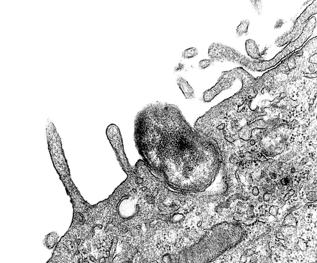
| Transmission Electron Microscope (TEM) image captured as the process of phagocytosis was underway. Here, you are able to see as an Orientia tsutsugamushi bacterium, formerly known as Rickettsia tsutsugamushi, was being ingested by a mouse peritoneal mesothelial cell. Note how the would-be host cell membrane had not yet entirely enveloped the bacterium. | |
| Lizenzart: | Lizenzpflichtig |
| Credit: | Science Photo Library / CDC / Science Source |
| Bildgröße: | 4050 px × 3362 px |
| Modell-Rechte: | nicht erforderlich |
| Eigentums-Rechte: | nicht erforderlich |
| Restrictions: | - |
Preise für dieses Bild ab 15 €
Universitäten & Organisationen
(Informationsmaterial Digital, Informationsmaterial Print, Lehrmaterial Digital etc.)
ab 15 €
Redaktionell
(Bücher, Bücher: Sach- und Fachliteratur, Digitale Medien (redaktionell) etc.)
ab 30 €
Werbung
(Anzeigen, Aussenwerbung, Digitale Medien, Fernsehwerbung, Karten, Werbemittel, Zeitschriften etc.)
ab 55 €
Handelsprodukte
(bedruckte Textilie, Kalender, Postkarte, Grußkarte, Verpackung etc.)
ab 75 €
Pauschalpreise
Rechtepakete für die unbeschränkte Bildnutzung in Print oder Online
ab 495 €
Keywords
- abnormal,
- Anomalie,
- bakteriell,
- Bakterien,
- Bakteriologie,
- Bakterium,
- Elektronenmikroskopie,
- elektronenmikroskopische Aufnahme,
- em,
- Erreger,
- gramnegativ,
- Histopathologie,
- histopathologisch,
- Kondition,
- krank,
- Krankheit,
- Mikrofotografie,
- Mikrographie,
- Mikroorganismus,
- Mikroskopie,
- mikroskopisch,
- mikroskopischer Organismus,
- Orientia tsutsugamushi,
- pathogen,
- Pathologie,
- pathologisch,
- Phagozytose,
- Prokaryot,
- Proteobakterien,
- Störung,
- SW,
- tem,
- Transmissionselektronenmikroskopie,
- transmissionselektronenmikroskopische Aufnahme,
- ungesund