Female pelvic arteries and organs, 1866 illustration
Bildnummer 12645175
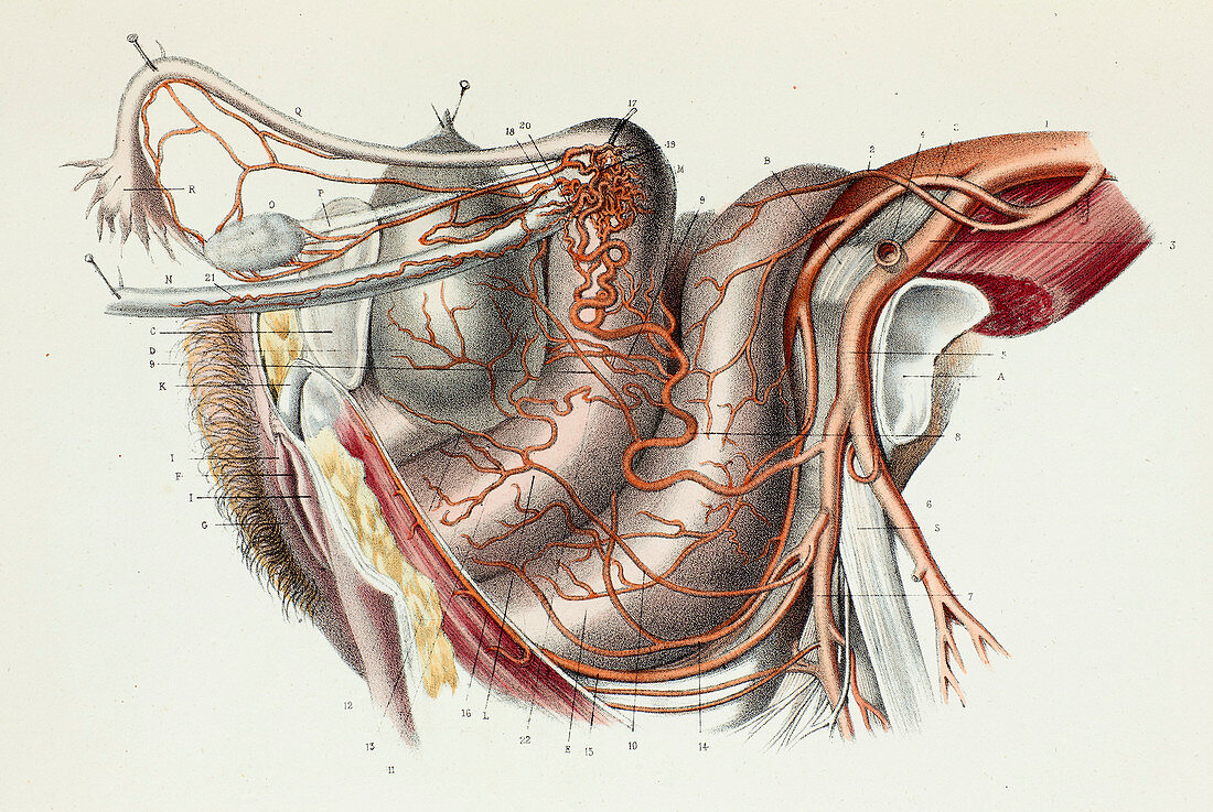
| Female pelvic arteries and organs, 1866 illustration. Lateral view of a dissection showing the arteries (red) supplying blood to the organs in a woman's pelvic region. From right to left, starting at centre, are the rectum, the uterus and vagina, the bladder, the pelvic bone and the external genitals. The fallopian tubes and ovaries are also shown. This page is plate 55 from the third volume of 'Atlas d'anatomie descriptive du corps humain' (1844-1866) by French anatomists Constantin Bonamy and Paul Broca. This work described the anatomy of the human body with over 250 hand-coloured lithographs. The illustrations were by Emile Beau, with the text by Bonamy and Broca. The three volumes were bound as four books in 1866 when the atlas was completed. This page is from the fourth book 'Generation Respiration', on the genitourinary and respiratory systems. | |
| Lizenzart: | Lizenzpflichtig |
| Credit: | Science Photo Library |
| Bildgröße: | 7156 px × 4792 px |
| Modell-Rechte: | nicht erforderlich |
| Eigentums-Rechte: | nicht erforderlich |
| Restrictions: | - |
Preise für dieses Bild ab 15 €
Universitäten & Organisationen
(Informationsmaterial Digital, Informationsmaterial Print, Lehrmaterial Digital etc.)
ab 15 €
Redaktionell
(Bücher, Bücher: Sach- und Fachliteratur, Digitale Medien (redaktionell) etc.)
ab 30 €
Werbung
(Anzeigen, Aussenwerbung, Digitale Medien, Fernsehwerbung, Karten, Werbemittel, Zeitschriften etc.)
ab 55 €
Handelsprodukte
(bedruckte Textilie, Kalender, Postkarte, Grußkarte, Verpackung etc.)
ab 75 €
Pauschalpreise
Rechtepakete für die unbeschränkte Bildnutzung in Print oder Online
ab 495 €
Keywords
- 1800er Jahre,
- 19. Jahrhundert,
- Anatomie,
- anatomisch,
- Arterie,
- arteriell,
- Arterien,
- Becken,
- Biologie,
- biologisch,
- Blase,
- Blatt,
- Blutgefäß,
- Blutgefäße,
- Buch,
- Eileiter,
- Enddarm,
- europäisch,
- Französisch,
- Frau,
- Geschichte,
- gesund,
- historisch,
- Illustration,
- Kreislauf,
- Kunstwerk,
- Lithographie,
- menschlicher Körper,
- Niemand,
- normal,
- Organ,
- pelvin,
- rektal,
- Reproduktion,
- Röhren,
- uterin,
- Uterus,
- Vagina,
- vaskulär,
- Veröffentlichung,
- weibliches Fortpflanzungssystem