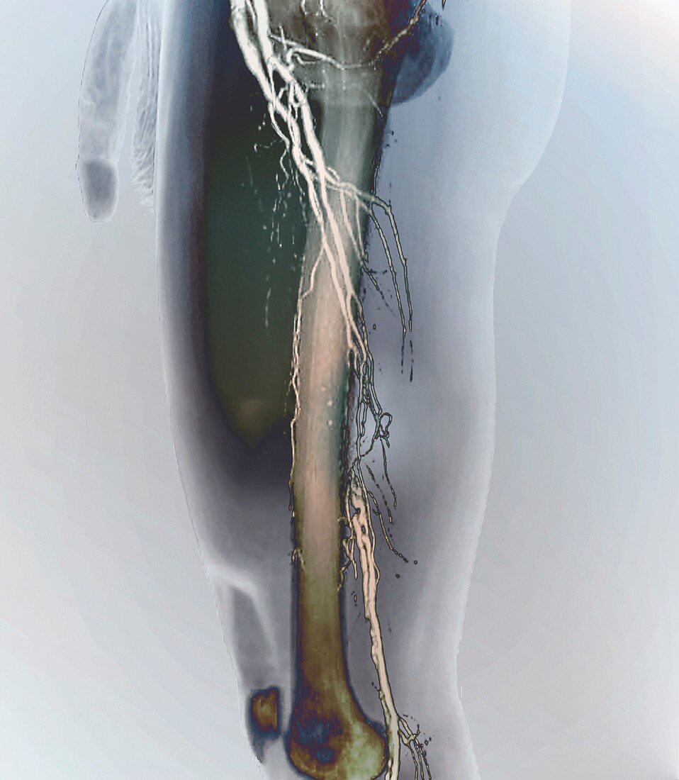
Nearly blocked femoral arteries, 3D CT angiogram
Bildnummer 12644430

| Nearly blocked femoral arteries. Coloured lateral 3D computed tomography (CT) angiogram (blood vessel X-ray) of the left leg of a 62-year-old man with preocclusive stenosis of the femoral arteries in his thigh. The arteries (white) have been highlighted using a contrast medium. Preocclusive stenosis is where a blood vessels has a narrowed area (stenosis) and is close to being completely blocked (occluded). In this case, collateral blood vessels in the rest of the leg have helped maintain the blood flow around the affected area (near centre). This lateral (side) view includes the patient's soft tissues and thigh bone (femur). | |
| Lizenzart: | Lizenzpflichtig |
| Credit: | Science Photo Library / Zephyr |
| Bildgröße: | 3902 px × 4479 px |
| Modell-Rechte: | nicht erforderlich |
| Eigentums-Rechte: | nicht erforderlich |
| Restrictions: | - |
Preise für dieses Bild ab 15 €
Universitäten & Organisationen
(Informationsmaterial Digital, Informationsmaterial Print, Lehrmaterial Digital etc.)
ab 15 €
Redaktionell
(Bücher, Bücher: Sach- und Fachliteratur, Digitale Medien (redaktionell) etc.)
ab 30 €
Werbung
(Anzeigen, Aussenwerbung, Digitale Medien, Fernsehwerbung, Karten, Werbemittel, Zeitschriften etc.)
ab 55 €
Handelsprodukte
(bedruckte Textilie, Kalender, Postkarte, Grußkarte, Verpackung etc.)
ab 75 €
Pauschalpreise
Rechtepakete für die unbeschränkte Bildnutzung in Print oder Online
ab 495 €
Keywords
- 3 dimensional,
- 3-d,
- 3-dimensional,
- 3D,
- 60er Jahre,
- abnormal,
- Angiografie,
- Angiogramm,
- Arterie,
- arteriell,
- Arterien,
- Bein,
- Blockierung,
- Blutgefäß,
- Blutgefäße,
- Computertomographie,
- CT-Scan,
- Dreidimensional,
- Erwachsene,
- farbig,
- Femoralarterie,
- geduldig,
- gefärbt,
- Kondition,
- Kontrastmittel,
- Krankheit,
- Kreislauf,
- Mann,
- Männlich,
- Medizin,
- medizinisch,
- menschlicher Körper,
- Niemand,
- Scanner,
- Schenkel,
- sechziger Jahre,
- Seitenansicht,
- seitlich,
- Stenose,
- Störung,
- ungesund,
- vaskulär,
- verkleinert,
- verstopft,
- weißer Hintergrund
