Subdural haematoma, 3D CT angiogram
Bildnummer 12644417
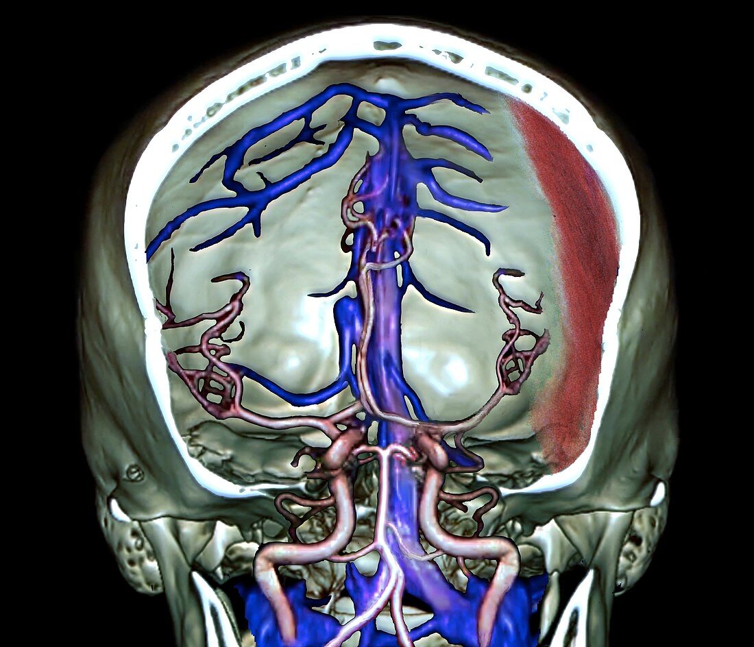
| Subdural haematoma. Coloured frontal 3D computed tomography (CT) angiogram, showing a cutaway view of the skull and brain of a 35-year-old man with a subdural haematoma (red, centre right) on the left side of his brain due to a car accident. The scan shows arteries (pink) and venous sinuses (blue). A haematoma is an accumulation of blood from internal bleeding, in this case into the space between the dura mater (outer brain membrane) and the brain. This subdural haematoma is affecting the left temporoparietal region of the brain. Typically caused by blunt force trauma to the skull, surgery may be required to relieve pressure caused by the internal bleeding. | |
| Lizenzart: | Lizenzpflichtig |
| Credit: | Science Photo Library / Zephyr |
| Bildgröße: | 4512 px × 3873 px |
| Modell-Rechte: | nicht erforderlich |
| Eigentums-Rechte: | nicht erforderlich |
| Restrictions: | - |
Preise für dieses Bild ab 15 €
Universitäten & Organisationen
(Informationsmaterial Digital, Informationsmaterial Print, Lehrmaterial Digital etc.)
ab 15 €
Redaktionell
(Bücher, Bücher: Sach- und Fachliteratur, Digitale Medien (redaktionell) etc.)
ab 30 €
Werbung
(Anzeigen, Aussenwerbung, Digitale Medien, Fernsehwerbung, Karten, Werbemittel, Zeitschriften etc.)
ab 55 €
Handelsprodukte
(bedruckte Textilie, Kalender, Postkarte, Grußkarte, Verpackung etc.)
ab 75 €
Pauschalpreise
Rechtepakete für die unbeschränkte Bildnutzung in Print oder Online
ab 495 €
Keywords
- 3 dimensional,
- 3-d,
- 3-dimensional,
- 30er Jahre,
- 3D,
- abnormal,
- Angiografie,
- Angiogramm,
- anterior,
- Arterie,
- Arterien,
- Autounfall,
- Blut,
- Blutung,
- cerebral,
- Computertomographie,
- CT-Scan,
- Cutaway,
- Diagnose,
- Dreidimensional,
- dreißiger Jahre,
- Erwachsene,
- farbig,
- Frontal,
- geduldig,
- gefärbt,
- Gehirn,
- Hämatom,
- Kondition,
- Kopf,
- Krankheit,
- Mann,
- Männlich,
- Medizin,
- medizinisch,
- menschlicher Körper,
- Neurologie,
- neurologisch,
- Niemand,
- Querschnitt,
- rta,
- Scanner,
- schwarzer Hintergrund,
- Sektion,
- sektioniert,
- Störung,
- ungesund,
- vaskulär,
- Verkehrsunfall,
- verletzt,
- Verletzung,
- Vorderansicht