Subacute infarct, MRI
Bildnummer 12641620
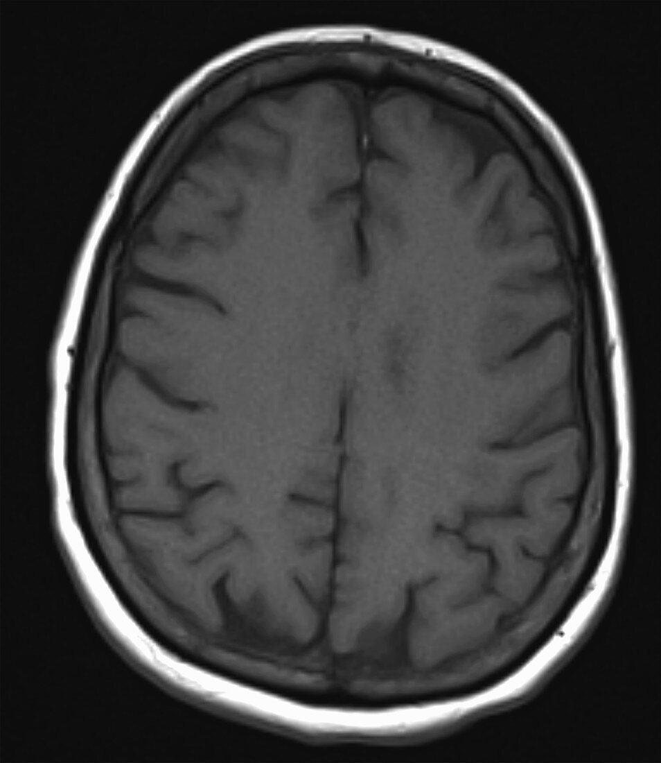
| Subacute infarct in a 62 year old female with an 8 day history of aphasia. Axial T1 1.5T MRI of the brain reveals intermediate intensity subcortical restricted EPI diffusion DWI signal in the left parietal lobe with relatively bright corresponding signal on the ADC map, bright FLAIR and T2 W signal, subtle low T1 signal pre contrast and post gadolinium enhancement. Signal changes are characteristic of a subacute infarct. Subcortical location indicates embolic source of stroke. | |
| Lizenzart: | Lizenzpflichtig |
| Credit: | Science Photo Library / Steven Needell |
| Bildgröße: | 2232 px × 2574 px |
| Modell-Rechte: | nicht erforderlich |
| Eigentums-Rechte: | nicht erforderlich |
| Restrictions: | - |
Preise für dieses Bild ab 15 €
Universitäten & Organisationen
(Informationsmaterial Digital, Informationsmaterial Print, Lehrmaterial Digital etc.)
ab 15 €
Redaktionell
(Bücher, Bücher: Sach- und Fachliteratur, Digitale Medien (redaktionell) etc.)
ab 30 €
Werbung
(Anzeigen, Aussenwerbung, Digitale Medien, Fernsehwerbung, Karten, Werbemittel, Zeitschriften etc.)
ab 55 €
Handelsprodukte
(bedruckte Textilie, Kalender, Postkarte, Grußkarte, Verpackung etc.)
ab 75 €
Pauschalpreise
Rechtepakete für die unbeschränkte Bildnutzung in Print oder Online
ab 495 €
Keywords
- abnormal,
- ADC,
- Akut,
- Attacke,
- Bildgebung,
- cerebral,
- chronisch,
- CVA,
- Diagnose,
- Flair,
- Gehirn,
- Gerinnsel,
- Kondition,
- Krankheit,
- Lappen,
- Magnet,
- Medizin,
- medizinisch,
- medizinische Bildgebung,
- medizinischer Scan,
- MRI,
- Pathologie,
- pathologisch,
- Radiologie,
- Resonanz,
- Schlaganfall,
- Störung,
- T1,
- t2,
- Thrombus,
- ungesund,
- vaskulär,
- Weiblich