Blastic femur metastases, X-ray
Bildnummer 12641292
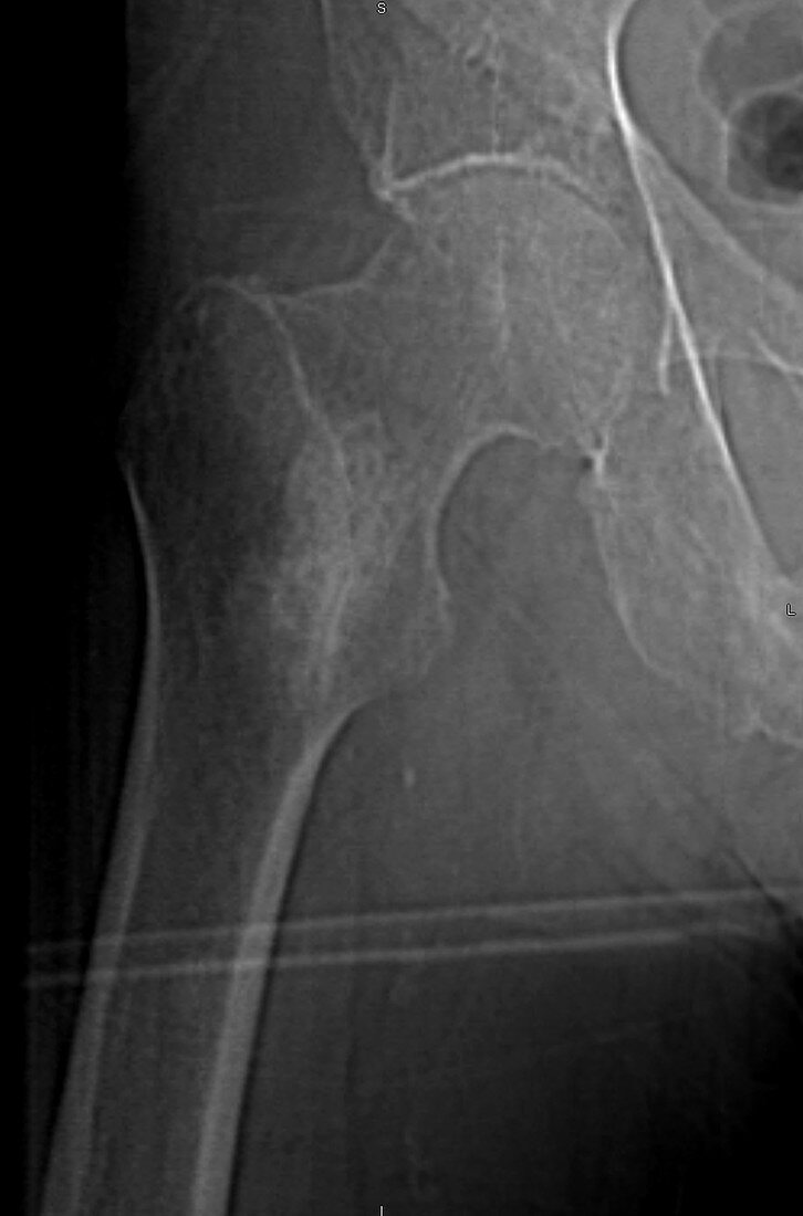
| AP femur X-ray from CT scanogram in a 90 year old male with prostate carcinoma reveals a 5 cm sclerotic high density lesion in the intertrochanteric proximal right femur, consistent with blastic metastatic prostate carcinoma. There is also a blastic metastasis in the inferior right pubic ramus. | |
| Lizenzart: | Lizenzpflichtig |
| Credit: | Science Photo Library / Steven Needell |
| Bildgröße: | 1614 px × 2446 px |
| Modell-Rechte: | nicht erforderlich |
| Eigentums-Rechte: | nicht erforderlich |
| Restrictions: | - |
Preise für dieses Bild ab 15 €
Universitäten & Organisationen
(Informationsmaterial Digital, Informationsmaterial Print, Lehrmaterial Digital etc.)
ab 15 €
Redaktionell
(Bücher, Bücher: Sach- und Fachliteratur, Digitale Medien (redaktionell) etc.)
ab 30 €
Werbung
(Anzeigen, Aussenwerbung, Digitale Medien, Fernsehwerbung, Karten, Werbemittel, Zeitschriften etc.)
ab 55 €
Handelsprodukte
(bedruckte Textilie, Kalender, Postkarte, Grußkarte, Verpackung etc.)
ab 75 €
Pauschalpreise
Rechtepakete für die unbeschränkte Bildnutzung in Print oder Online
ab 495 €
Keywords
- Alt,
- älter,
- Becken,
- Bildgebung,
- Computertomographie,
- ct,
- CT-Scan,
- Diagnose,
- Dichte,
- Femur,
- Frontal,
- hoch,
- Hüfte,
- Karzinom,
- Knochen,
- Kondition,
- Krankheit,
- Krebs,
- Männlich,
- Masse,
- Medizin,
- medizinisch,
- medizinische Bildgebung,
- medizinischer Scan,
- muskuloskeletal,
- normal,
- Pathologie,
- pathologisch,
- Prostata,
- Radiographie,
- Radiologie,
- Recht,
- Röntgen,
- Röntgenbild,
- Scan,
- Schenkel,
- Störung,
- Tumor,
- ungesund,
- Wunde