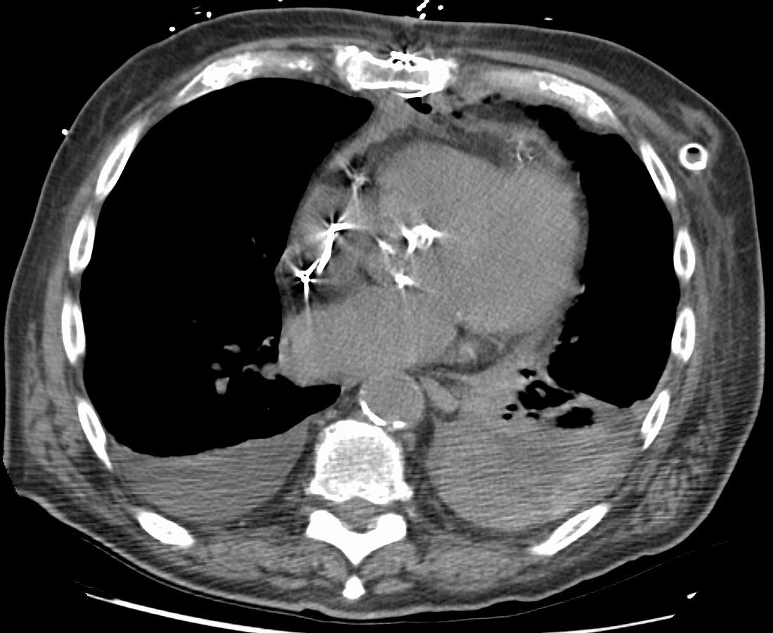
Hemothorax post cardiac surgery, CT scan
Bildnummer 12637970

| CT scan of an 83 year old male post cardiac surgery. This noncontrast volume axial CT image reveals bilateral pleural effusions. The fluid on the left has higher, heterogeneous density compared to the fluid on the right, consistent with haemorrhage and hemothorax. Left lower lobe atelectasis is present. There is a small left sided pneumothorax and an anterior left sided chest tube from recent surgery. | |
| Lizenzart: | Lizenzpflichtig |
| Credit: | Science Photo Library / Steven Needell |
| Bildgröße: | 2750 px × 2250 px |
| Modell-Rechte: | nicht erforderlich |
| Eigentums-Rechte: | nicht erforderlich |
| Restrictions: | - |
Preise für dieses Bild ab 15 €
Universitäten & Organisationen
(Informationsmaterial Digital, Informationsmaterial Print, Lehrmaterial Digital etc.)
ab 15 €
Redaktionell
(Bücher, Bücher: Sach- und Fachliteratur, Digitale Medien (redaktionell) etc.)
ab 30 €
Werbung
(Anzeigen, Aussenwerbung, Digitale Medien, Fernsehwerbung, Karten, Werbemittel, Zeitschriften etc.)
ab 55 €
Handelsprodukte
(bedruckte Textilie, Kalender, Postkarte, Grußkarte, Verpackung etc.)
ab 75 €
Pauschalpreise
Rechtepakete für die unbeschränkte Bildnutzung in Print oder Online
ab 495 €
Keywords
- abnormal,
- axial,
- Bild,
- Bildgebung,
- Blut,
- Blutung,
- Computertomographie,
- ct,
- CT-Scan,
- Diagnose,
- diagnostische Bildgebung,
- Dichte,
- Fluid,
- Kondition,
- Krankheit,
- links,
- Medizin,
- medizinisch,
- medizinische Bildgebung,
- medizinischer Scan,
- OP-Nachsorge,
- Pathologie,
- pathologisch,
- Recht,
- Scan,
- Störung,
- Truhe,
- Tube,
- ungesund,
- Volumen,
- Zusammenbruch
