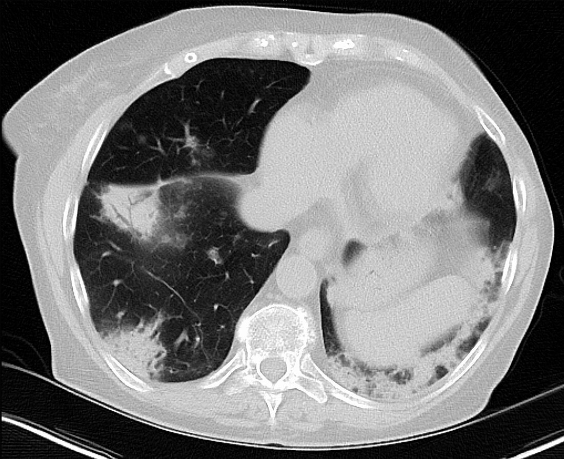
Eosinophilic pneumonia, CT scan
Bildnummer 12637965

| CT scan of the chest in a 70 year old female with chronic eosinophilic pneumonia (CEP). The scan reveals waxing and waning, fleeting pulmonary infiltrates showing non-segmental areas of airspace consolidation with peripheral predominance, pulmonary nodules, ground-glass opacities and linear band like opacities parallel to the pleural surface. Differential diagnosis includes eosinophilic pneumonia, Loeffler syndrome, vasculitis, and allergic bronchopulmonary aspergillosis and cryptogenic organizing pneumonia (COP), the idiopathic form of organizing pneumonia (formerly called bronchiolitis obliterans organizing pneumonia or BOOP). | |
| Lizenzart: | Lizenzpflichtig |
| Credit: | Science Photo Library / Steven Needell |
| Bildgröße: | 2752 px × 2240 px |
| Modell-Rechte: | nicht erforderlich |
| Eigentums-Rechte: | nicht erforderlich |
| Restrictions: | - |
Preise für dieses Bild ab 15 €
Universitäten & Organisationen
(Informationsmaterial Digital, Informationsmaterial Print, Lehrmaterial Digital etc.)
ab 15 €
Redaktionell
(Bücher, Bücher: Sach- und Fachliteratur, Digitale Medien (redaktionell) etc.)
ab 30 €
Werbung
(Anzeigen, Aussenwerbung, Digitale Medien, Fernsehwerbung, Karten, Werbemittel, Zeitschriften etc.)
ab 55 €
Handelsprodukte
(bedruckte Textilie, Kalender, Postkarte, Grußkarte, Verpackung etc.)
ab 75 €
Pauschalpreise
Rechtepakete für die unbeschränkte Bildnutzung in Print oder Online
ab 495 €
Keywords
- abnormal,
- Akut,
- allergisch,
- Bildgebung,
- Boden,
- chronisch,
- Computertomographie,
- ct,
- CT-Scan,
- Diagnose,
- diagnostische Bildgebung,
- Glas,
- Kondition,
- Krankheit,
- Linear,
- Luftraum,
- Lungenentzündung,
- Medizin,
- medizinisch,
- medizinische Bildgebung,
- medizinischer Scan,
- Oberfläche,
- Opazität,
- Parallel,
- Pathologie,
- pathologisch,
- peripher,
- pulmonal,
- Röntgen,
- Röntgenbild,
- Scan,
- Störung,
- Syndrom,
- Thorax,
- Truhe,
- ungesund,
- Weiblich
