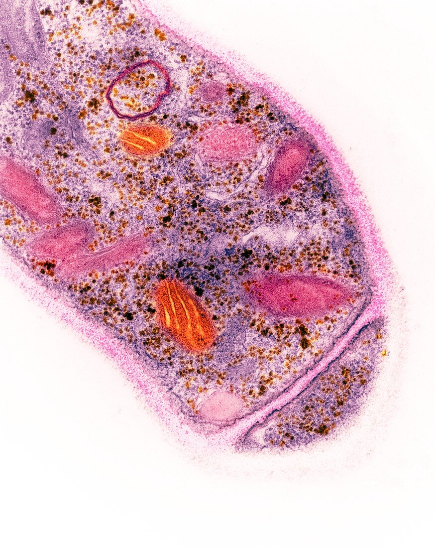
Trichoderma reesei fungus, TEM
Bildnummer 12539241

| Coloured transmission electron micrograph (TEM) of a section through a hypha (vegetative filament) of a Trichoderma reesei fungus. Trichoderma is a soil fungus involved in the decomposition of plant material. Surrounding the hypha is a thick cell wall (pink and white). At lower right is a septum (dividing wall) between two hyphal cells. In the fungal cell above the septum certain organelles can be seen. Mitochondria, the sites for energy production in the cell, are orange. Ribosomes, the sites of protein synthesis, are visible as small dark dots. Magnification x31, 500 when printed at 10 centimetres tall. | |
| Lizenzart: | Lizenzpflichtig |
| Credit: | Science Photo Library / Lounatmaa, Dr. Kari |
| Bildgröße: | 3733 px × 4700 px |
| Modell-Rechte: | nicht erforderlich |
| Eigentums-Rechte: | nicht erforderlich |
| Restrictions: | - |
Preise für dieses Bild ab 15 €
Universitäten & Organisationen
(Informationsmaterial Digital, Informationsmaterial Print, Lehrmaterial Digital etc.)
ab 15 €
Redaktionell
(Bücher, Bücher: Sach- und Fachliteratur, Digitale Medien (redaktionell) etc.)
ab 30 €
Werbung
(Anzeigen, Aussenwerbung, Digitale Medien, Fernsehwerbung, Karten, Werbemittel, Zeitschriften etc.)
ab 55 €
Handelsprodukte
(bedruckte Textilie, Kalender, Postkarte, Grußkarte, Verpackung etc.)
ab 75 €
Pauschalpreise
Rechtepakete für die unbeschränkte Bildnutzung in Print oder Online
ab 495 €
Keywords
- Anatomie,
- Art,
- elektronenmikroskopische Aufnahme,
- Eumycota,
- farbig,
- gefärbt,
- Hyphe,
- Mikrobiologie,
- mikrobiologisch,
- Mitochondrien,
- Natur,
- Niemand,
- Organellen,
- Pilz,
- Pilz-,
- Pilze,
- Pilzkunde,
- tem,
- transmissionselektronenmikroskopische Aufnahme,
- Übertragung,
- weißer Hintergrund,
- Zellenwand,
- Zellstruktur
