Dentine tooth tissue, SEM
Bildnummer 12528254
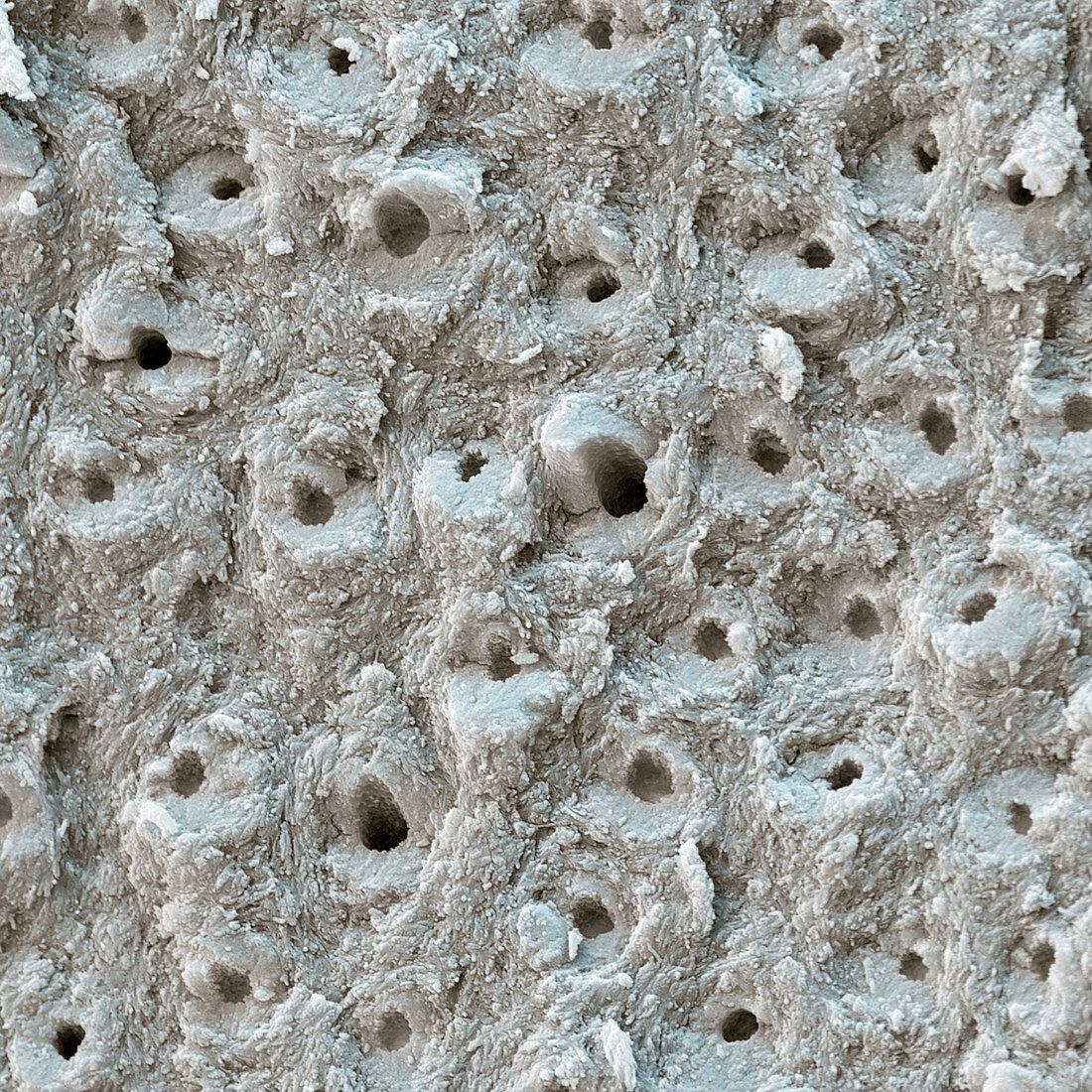
| Dentine tooth tissue. Coloured scanning electron micrograph (SEM) of dentine (substantia eburnea), which is a mineralised connective tissue found under a tooth's enamel. It forms the bulk of a tooth and differs from bone in its microscopic structure, which is seen here. Shown in cross section are the dentinal tubules (dental canaliculi), which are where extensions from the odontoblast cells (part of the pulp at the core of a tooth) have formed the surrounding dentine matrix. These odontoblast extensions (or processes) allow dentine to rebuild itself, unlike enamel. Magnification: x2500 when printed 10 centimetres wide. | |
| Lizenzart: | Lizenzpflichtig |
| Credit: | Science Photo Library / EYE OF SCIENCE |
| Bildgröße: | 4000 px × 4000 px |
| Modell-Rechte: | nicht erforderlich |
| Eigentums-Rechte: | nicht erforderlich |
| Restrictions: |
|
Preise für dieses Bild ab 15 €
Universitäten & Organisationen
(Informationsmaterial Digital, Informationsmaterial Print, Lehrmaterial Digital etc.)
ab 15 €
Redaktionell
(Bücher, Bücher: Sach- und Fachliteratur, Digitale Medien (redaktionell) etc.)
ab 30 €
Werbung
(Anzeigen, Aussenwerbung, Digitale Medien, Fernsehwerbung, Karten, Werbemittel, Zeitschriften etc.)
ab 55 €
Handelsprodukte
(bedruckte Textilie, Kalender, Postkarte, Grußkarte, Verpackung etc.)
ab 75 €
Pauschalpreise
Rechtepakete für die unbeschränkte Bildnutzung in Print oder Online
ab 495 €