Cerebellar glomerulus, TEM
Bildnummer 12504718
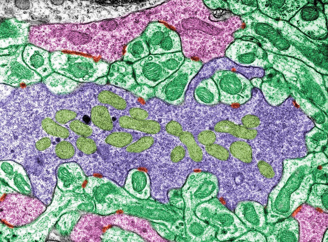
| Coloured transmission electron micrograph (TEM) of a section through a cerebellar glomerulus. Across centre is a mossy fiber (purple), one of the inputs to the cerebellum. Within it are synaptic vesicles and mitochondria (khaki). It is surrounded by many granule cell dendrites (green) and, outwardly, axon terminals of Golgi II neurons (pink). The synaptic contacts are labelled in red. | |
| Lizenzart: | Lizenzpflichtig |
| Credit: | Science Photo Library / JOSE CALVO |
| Bildgröße: | 3771 px × 2786 px |
| Modell-Rechte: | nicht erforderlich |
| Eigentums-Rechte: | nicht erforderlich |
| Restrictions: | - |
Preise für dieses Bild ab 15 €
Universitäten & Organisationen
(Informationsmaterial Digital, Informationsmaterial Print, Lehrmaterial Digital etc.)
ab 15 €
Redaktionell
(Bücher, Bücher: Sach- und Fachliteratur, Digitale Medien (redaktionell) etc.)
ab 30 €
Werbung
(Anzeigen, Aussenwerbung, Digitale Medien, Fernsehwerbung, Karten, Werbemittel, Zeitschriften etc.)
ab 55 €
Handelsprodukte
(bedruckte Textilie, Kalender, Postkarte, Grußkarte, Verpackung etc.)
ab 75 €
Pauschalpreise
Rechtepakete für die unbeschränkte Bildnutzung in Print oder Online
ab 495 €
Keywords
- Biologie,
- biologisch,
- Bürstensaum,
- Dendrit,
- farbig,
- Fehlfarbe,
- gefärbt,
- Glomerulus,
- Histologie,
- histologisch,
- Kleinhirn,
- Mikroskopie,
- Neuron,
- Niemand,
- Synapse,
- tem,
- Transmissionselektronenmikroskop,
- transmissionselektronenmikroskopische Aufnahme,
- Ultrastruktur,
- Zelle,
- zentrales Nervensystem,
- Zytologie,
- Zytologisch