Coloured PET brain scan during listening exercise
Bildnummer 12496932
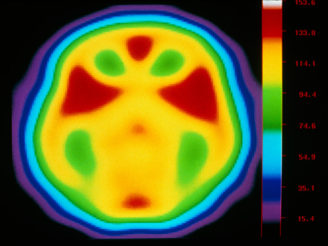
| Brain's auditory centre. Coloured positron emission tomography (PET) scan of the brain during a listening exercise. The exercise involved unfocused listening to background noise. In this axial (horizontal) scan the front of the brain is at top. The scan shows oxygen and water levels from low (blue) to high (red). These levels correspond to brain activity, the highest levels being in the auditory cortex (red triangles) of the two temporal lobes. PET scans use radioactively-labelled substances introduced into the blood to view metabolic activity. See P335/035 for a PET scan taken during focused listening. | |
| Lizenzart: | Lizenzpflichtig |
| Credit: | Science Photo Library / MONTREAL NEUROLOGICAL INSTITUTE |
| Bildgröße: | 4367 px × 3279 px |
| Modell-Rechte: | nicht erforderlich |
| Eigentums-Rechte: | nicht erforderlich |
| Restrictions: | - |
Preise für dieses Bild ab 15 €
Universitäten & Organisationen
(Informationsmaterial Digital, Informationsmaterial Print, Lehrmaterial Digital etc.)
ab 15 €
Redaktionell
(Bücher, Bücher: Sach- und Fachliteratur, Digitale Medien (redaktionell) etc.)
ab 30 €
Werbung
(Anzeigen, Aussenwerbung, Digitale Medien, Fernsehwerbung, Karten, Werbemittel, Zeitschriften etc.)
ab 55 €
Handelsprodukte
(bedruckte Textilie, Kalender, Postkarte, Grußkarte, Verpackung etc.)
ab 75 €
Pauschalpreise
Rechtepakete für die unbeschränkte Bildnutzung in Print oder Online
ab 495 €