Mouse embryo heart, SEM
Bildnummer 12445189
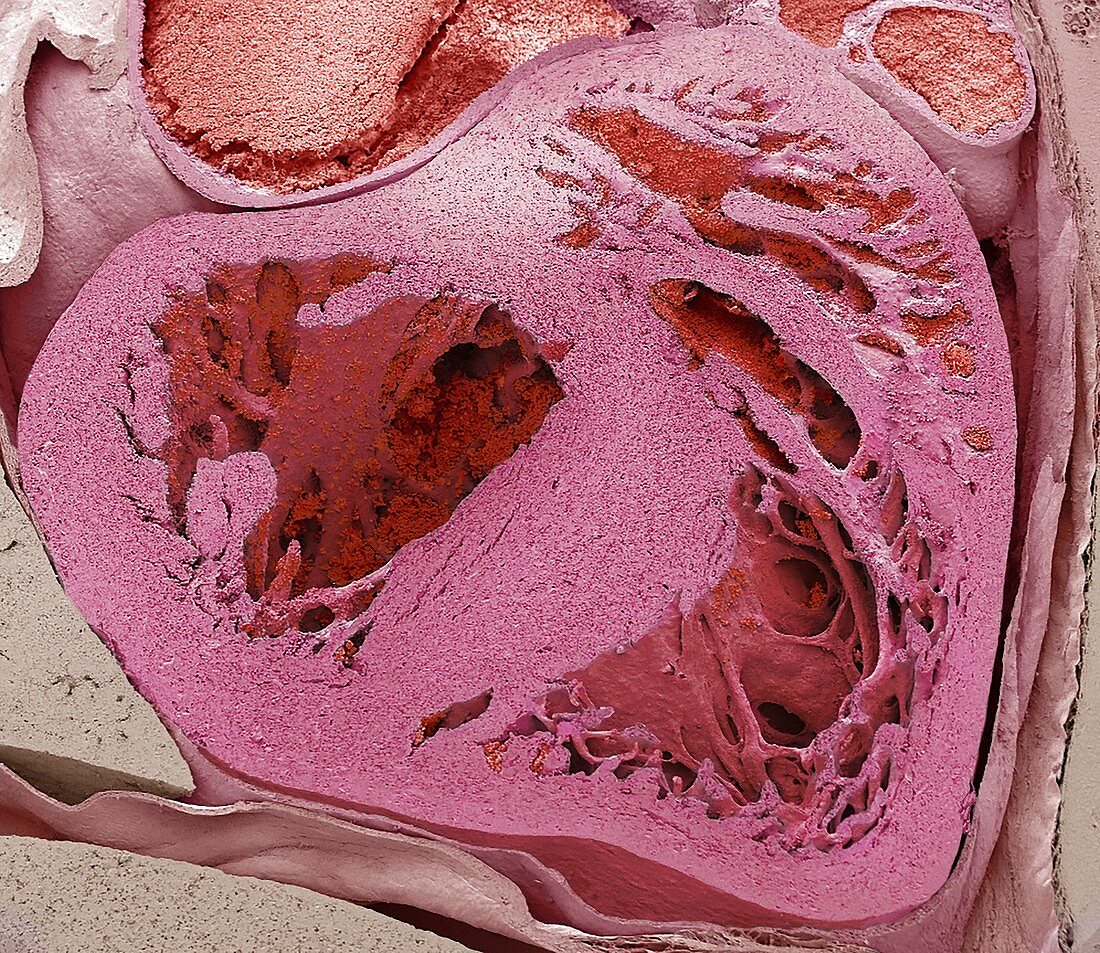
| Mouse embryo heart. Coloured scanning electron micrograph (SEM) of a sagittal (vertical and central) section that has halved a mouse embryo heart. On the bottom left, the right ventricle and right atrium (partial top right) receive deoxygenated blood from the body through the superior and inferior vena cava. From the right ventricle the blood flows through the pulmonary arteries to the lungs. Oxygenated blood returns to the left atrium and ventricle (right) and is distributed to the body through the ascending aorta. Chordae tendineae are cord-like tendons that connect the papillary muscles to the tricuspid valve and the mitral valve in the heart and are visible in both ventricles. Mice breed rapidly, with a short gestation period of a few weeks, producing a litter of 3-12 young that are born naked and blind. Magnification: x60 when printed 10cm wide. | |
| Lizenzart: | Lizenzpflichtig |
| Credit: | Science Photo Library / Gschmeissner, Steve |
| Bildgröße: | 4572 px × 3962 px |
| Modell-Rechte: | nicht erforderlich |
| Eigentums-Rechte: | nicht erforderlich |
| Restrictions: | - |
Preise für dieses Bild ab 15 €
Universitäten & Organisationen
(Informationsmaterial Digital, Informationsmaterial Print, Lehrmaterial Digital etc.)
ab 15 €
Redaktionell
(Bücher, Bücher: Sach- und Fachliteratur, Digitale Medien (redaktionell) etc.)
ab 30 €
Werbung
(Anzeigen, Aussenwerbung, Digitale Medien, Fernsehwerbung, Karten, Werbemittel, Zeitschriften etc.)
ab 55 €
Handelsprodukte
(bedruckte Textilie, Kalender, Postkarte, Grußkarte, Verpackung etc.)
ab 75 €
Pauschalpreise
Rechtepakete für die unbeschränkte Bildnutzung in Print oder Online
ab 495 €
Keywords
- Anatomie,
- anatomisch,
- Arterien,
- Atrium,
- Biologie,
- biologisch,
- Blut,
- Embryo,
- Embryologie,
- Entwicklung,
- Farbig,
- Fauna,
- Fötus,
- gefärbt,
- geschnitten,
- halb,
- Herz,
- Histologie,
- histologisch,
- innere Organe,
- Maus,
- pulmonal,
- rasterelektronenmikroskopische Aufnahme,
- REM,
- Rückgrat,
- Säugetier,
- Scheibe,
- Sektion,
- sektioniert,
- Tierkörper,
- Vena-cava,
- Ventrikel