Rosemary leaf cross section (Rosmarinus officinalis), SEM
Bildnummer 12301516
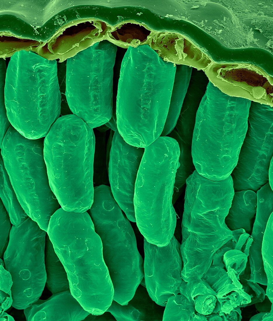
| Rosemary leaf cross section (Rosmarinus officinalis), coloured scanning electron micrograph (SEM). A thin waxy cuticle is present on the upper and lower surface of the leaf. The rectangular line of cells near top of the leaf (right below the cuticle) is the leaf epidermis containing epidermal cells (without chloroplasts). The cuticle and epidermis helps to protect the leaf. The leaf interior contains mesophyll parenchyma cells that consist of two types 1) palisade mesophyll, oblong cells near the upper epidermis arranged perpendicular to the epidermal cells and 2) spongy mesophyll, (not visible in this image). The packed palisade mesophyll cells contain chloroplasts that are the major leaf sites for photosynthesis. The spongy mesophyll cells are loosely packed, with few chloroplasts and are more involved with gas exchange. Magnification: x220 when shortest axis printed at 25 millimetres. | |
| Lizenzart: | Lizenzpflichtig |
| Credit: | Science Photo Library / DENNIS KUNKEL MICROSCOPY |
| Bildgröße: | 2726 px × 3206 px |
| Modell-Rechte: | nicht erforderlich |
| Eigentums-Rechte: | nicht erforderlich |
| Restrictions: | - |
Preise für dieses Bild ab 15 €
Universitäten & Organisationen
(Informationsmaterial Digital, Informationsmaterial Print, Lehrmaterial Digital etc.)
ab 15 €
Redaktionell
(Bücher, Bücher: Sach- und Fachliteratur, Digitale Medien (redaktionell) etc.)
ab 30 €
Werbung
(Anzeigen, Aussenwerbung, Digitale Medien, Fernsehwerbung, Karten, Werbemittel, Zeitschriften etc.)
ab 55 €
Handelsprodukte
(bedruckte Textilie, Kalender, Postkarte, Grußkarte, Verpackung etc.)
ab 75 €
Pauschalpreise
Rechtepakete für die unbeschränkte Bildnutzung in Print oder Online
ab 495 €