Euglena gracilis, TEM
Bildnummer 12298605
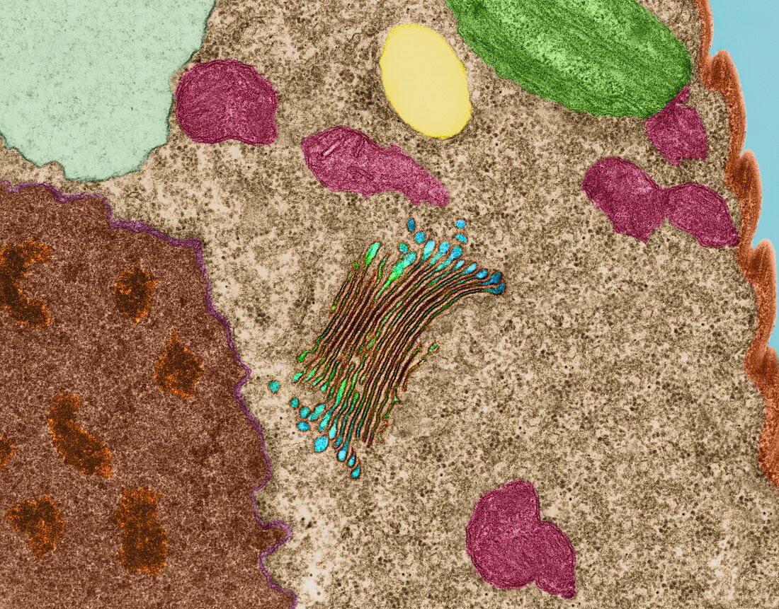
| Coloured scanning electron micrograph (TEM) of Euglena gracilis cell pellicle and internal cytoplasmic organelles: golgi apparatus (light green and blue), chloroplast (green), mitochondria (dark pink), pyrenoid body (light yellow), ribosomes (brown), vacuole (light blue-green), nuclear membrane (purple), nucleus (dark brown), chromatin (orange). Euglena spp. can change their shape readily due to microtubules located beneath their cell membrane (pellicle, light orange). Euglena gracilis is a fresh water, flagellated protozoan often classified in a group called euglenoids. Euglena spp. normally contain chloroplasts but in prolonged darkness they become heterotrophic and engulf other small organisms. Magnification: x5, 505 when shortest axis printed at 25 millimetres. | |
| Lizenzart: | Lizenzpflichtig |
| Credit: | Science Photo Library / DENNIS KUNKEL MICROSCOPY |
| Bildgröße: | 4218 px × 3300 px |
| Modell-Rechte: | nicht erforderlich |
| Eigentums-Rechte: | nicht erforderlich |
| Restrictions: | - |
Preise für dieses Bild ab 15 €
Universitäten & Organisationen
(Informationsmaterial Digital, Informationsmaterial Print, Lehrmaterial Digital etc.)
ab 15 €
Redaktionell
(Bücher, Bücher: Sach- und Fachliteratur, Digitale Medien (redaktionell) etc.)
ab 30 €
Werbung
(Anzeigen, Aussenwerbung, Digitale Medien, Fernsehwerbung, Karten, Werbemittel, Zeitschriften etc.)
ab 55 €
Handelsprodukte
(bedruckte Textilie, Kalender, Postkarte, Grußkarte, Verpackung etc.)
ab 75 €
Pauschalpreise
Rechtepakete für die unbeschränkte Bildnutzung in Print oder Online
ab 495 €
Keywords
- Alge,
- Algen,
- Atomkern,
- Briefumschlag,
- Chloroplasten,
- DNA,
- Elektron,
- Eukaryot,
- farbig,
- gefärbt,
- Gerät,
- Grün,
- Körper,
- Lipid,
- Membran,
- Mikrofotografie,
- Mikrotubuli,
- Mitochondrien,
- Mitochondrion,
- nuklear,
- Protist,
- Protista,
- Protozoen,
- Protozoon,
- Süßwasser,
- tem,
- thylakoid,
- Thylakoide,
- Übertragung,
- Wasser-,
- Zytoplasma