8 week embryonic Urinary System
Bildnummer 12071251
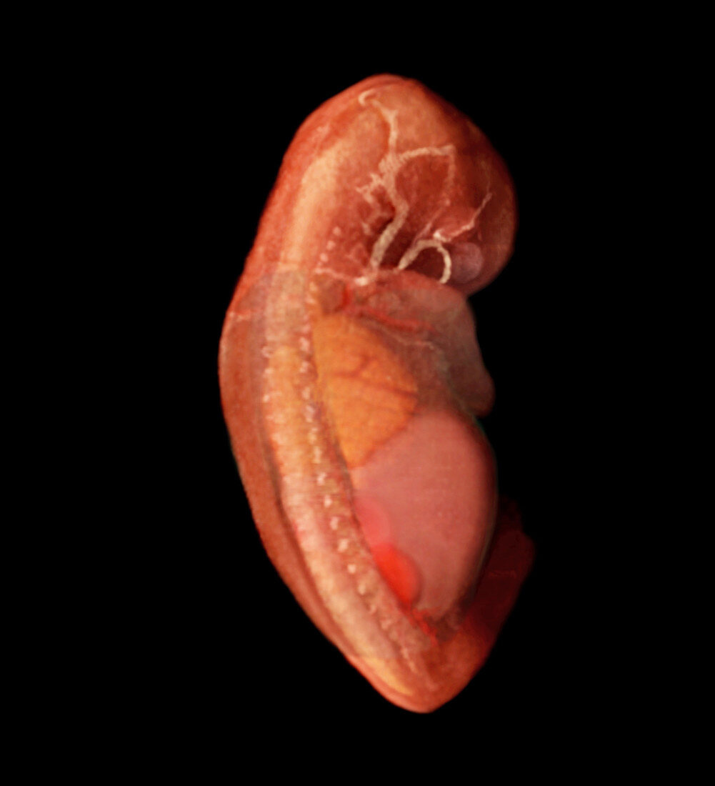
| Three-dimensional visualisation based on segmented human data from MRI scans of the internal anatomy of a fifty-five-day-old human embryo. The lungs with clearly defined lobes can be seen in yellow,the lighter coloured circulatory vessels can be seen feeding and draining waste from the tissues in her head,and the developing spinal column and enlarged liver are visible. At this stage in development,the liver is abnormally large due to its functioning to create blood cells | |
| Lizenzart: | Lizenzpflichtig |
| Credit: | Science Photo Library / Anatomical Travelogue |
| Bildgröße: | 2293 px × 2521 px |
| Modell-Rechte: | nicht erforderlich |
| Eigentums-Rechte: | nicht erforderlich |
| Restrictions: |
|
Preise für dieses Bild ab 15 €
Universitäten & Organisationen
(Informationsmaterial Digital, Informationsmaterial Print, Lehrmaterial Digital etc.)
ab 15 €
Redaktionell
(Bücher, Bücher: Sach- und Fachliteratur, Digitale Medien (redaktionell) etc.)
ab 30 €
Werbung
(Anzeigen, Aussenwerbung, Digitale Medien, Fernsehwerbung, Karten, Werbemittel, Zeitschriften etc.)
ab 55 €
Handelsprodukte
(bedruckte Textilie, Kalender, Postkarte, Grußkarte, Verpackung etc.)
ab 75 €
Pauschalpreise
Rechtepakete für die unbeschränkte Bildnutzung in Print oder Online
ab 495 €