Cell division, light micrograph
Bildnummer 12071195
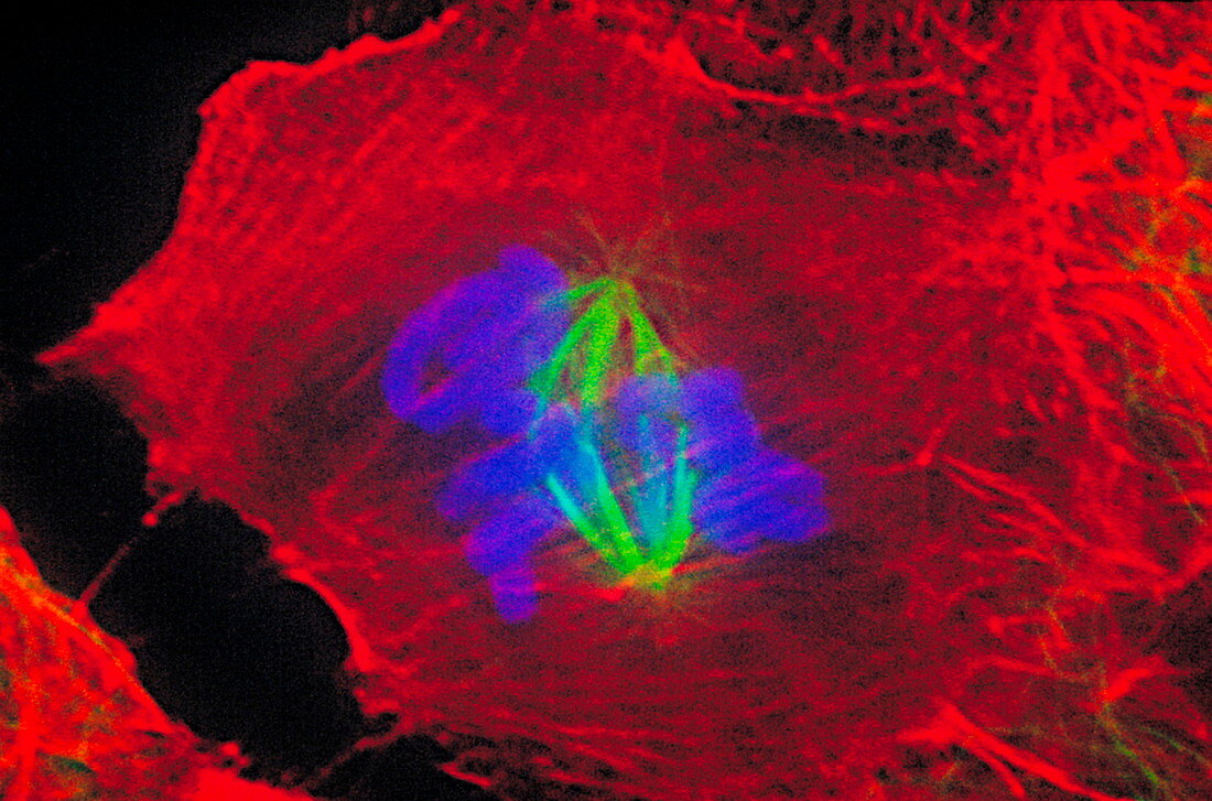
| Cell division. Immunofluorescent light micrograph of the prometaphase stage of cell division (mitosis). Antibodies have beeen used to attach fluorescent dyes to specific cell tissues. The nuclear membrane (not seen) that encloses the chromosomes (blue) is breaking down. This allows the chromosomes contained within it to replicate and form two new nuclei. The actin microfilaments (red) and tubulin microtubules (green) of the cytoskeleton maintain the structure of the cell. Mitotic cell division produces two identical daughter cells. Cell taken from a rat kangaroo kidney. | |
| Lizenzart: | Lizenzpflichtig |
| Credit: | Science Photo Library / Waters, Jennifer |
| Bildgröße: | 3767 px × 2489 px |
| Modell-Rechte: | nicht erforderlich |
| Eigentums-Rechte: | nicht erforderlich |
| Restrictions: |
|
Preise für dieses Bild ab 15 €
Universitäten & Organisationen
(Informationsmaterial Digital, Informationsmaterial Print, Lehrmaterial Digital etc.)
ab 15 €
Redaktionell
(Bücher, Bücher: Sach- und Fachliteratur, Digitale Medien (redaktionell) etc.)
ab 30 €
Werbung
(Anzeigen, Aussenwerbung, Digitale Medien, Fernsehwerbung, Karten, Werbemittel, Zeitschriften etc.)
ab 55 €
Handelsprodukte
(bedruckte Textilie, Kalender, Postkarte, Grußkarte, Verpackung etc.)
ab 75 €
Pauschalpreise
Rechtepakete für die unbeschränkte Bildnutzung in Print oder Online
ab 495 €