Human tooth,light micrograph
Bildnummer 12052679
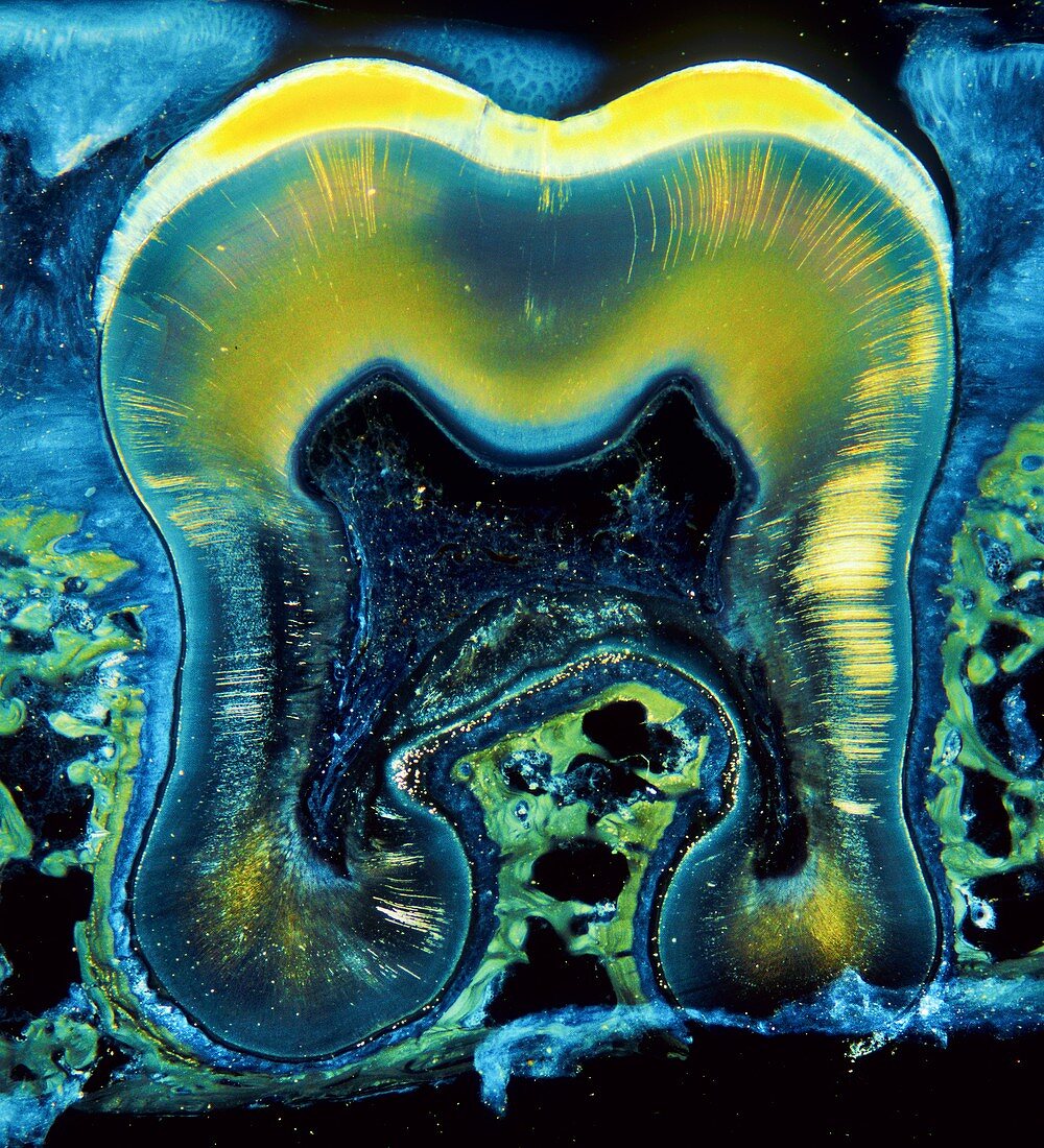
| Darkfield microscopy of a vertical section of a molar tooth. The specimen is an unstained thick section which displays colours due to the refraction or scattering of light. Enamel is bright gold,the deeper crown and roots showing in part the bony dentinal tubules with the central black core representing the pulp cavity for vessels and nerves. The green-tinted honeycomb-type structures are jaw bone. Between the bone and the tooth the periodontal ligament shows a blue colour. Magnification x 4.5 when printed at 10 cm | |
| Lizenzart: | Lizenzpflichtig |
| Credit: | Science Photo Library / Microscape |
| Bildgröße: | 4091 px × 4500 px |
| Modell-Rechte: | nicht erforderlich |
| Eigentums-Rechte: | nicht erforderlich |
| Restrictions: | - |
Preise für dieses Bild ab 15 €
Universitäten & Organisationen
(Informationsmaterial Digital, Informationsmaterial Print, Lehrmaterial Digital etc.)
ab 15 €
Redaktionell
(Bücher, Bücher: Sach- und Fachliteratur, Digitale Medien (redaktionell) etc.)
ab 30 €
Werbung
(Anzeigen, Aussenwerbung, Digitale Medien, Fernsehwerbung, Karten, Werbemittel, Zeitschriften etc.)
ab 55 €
Handelsprodukte
(bedruckte Textilie, Kalender, Postkarte, Grußkarte, Verpackung etc.)
ab 75 €
Pauschalpreise
Rechtepakete für die unbeschränkte Bildnutzung in Print oder Online
ab 495 €