MRI of Normal and Injured Brains
Bildnummer 12037125
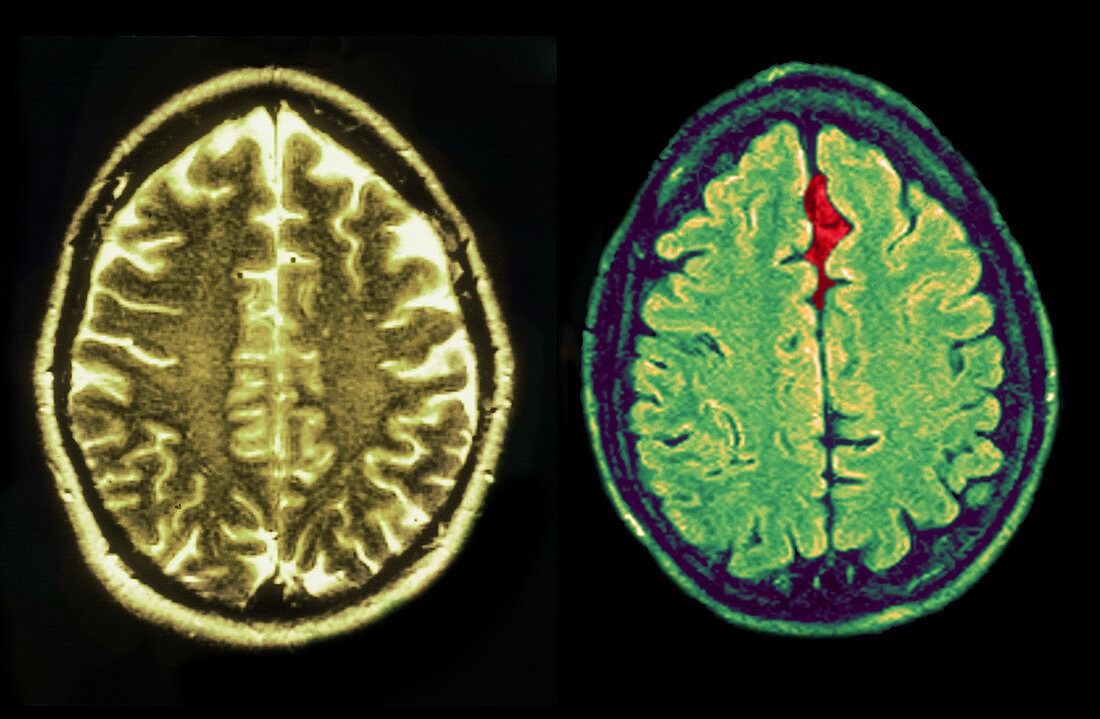
| On the left is an MRI scan (T2 weighted,axial view) of the normal brain of a 54-year-old female. On the right is an axial MRI of the brain of a 26-year-old male whose head was injured in a car accident. Diagnosis from the MRI showed a small arachnoid cyst in the parasagittal anterior left frontal region (red). All other aspects were normal | |
| Lizenzart: | Lizenzpflichtig |
| Credit: | Science Photo Library / Wilson, Jessica |
| Bildgröße: | 4922 px × 3218 px |
| Modell-Rechte: | nicht erforderlich |
| Eigentums-Rechte: | nicht erforderlich |
| Restrictions: |
|
Preise für dieses Bild ab 15 €
Universitäten & Organisationen
(Informationsmaterial Digital, Informationsmaterial Print, Lehrmaterial Digital etc.)
ab 15 €
Redaktionell
(Bücher, Bücher: Sach- und Fachliteratur, Digitale Medien (redaktionell) etc.)
ab 30 €
Werbung
(Anzeigen, Aussenwerbung, Digitale Medien, Fernsehwerbung, Karten, Werbemittel, Zeitschriften etc.)
ab 55 €
Handelsprodukte
(bedruckte Textilie, Kalender, Postkarte, Grußkarte, Verpackung etc.)
ab 75 €
Pauschalpreise
Rechtepakete für die unbeschränkte Bildnutzung in Print oder Online
ab 495 €
Keywords
- abnormal,
- Arachnoidalzyste,
- Atrium,
- Autounfall,
- csf,
- Diagnose,
- Einschlag,
- Erweiterung,
- Farbe,
- Gehirn,
- gutartig,
- Kontrast,
- Krankheit,
- Magnetresonanztomografie,
- medizinisch,
- medizinische Bildgebung,
- MRI,
- Nervensystem,
- Neuroimaging,
- normal,
- Pathologie,
- Scan,
- Ventrikel,
- verbessert,
- Vergleich,
- verletzt,
- zentrales Nervensystem,
- Zusammengesetzt,
- Zyste