MRI of Normal Brain and Hemispherectomy
Bildnummer 12037121
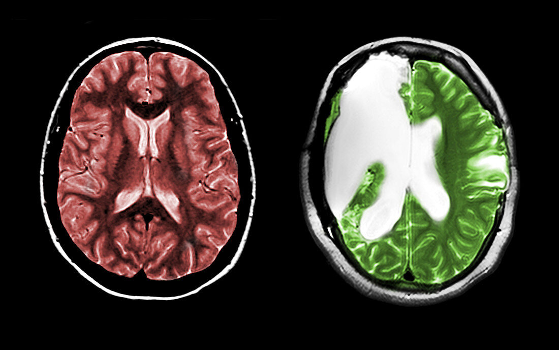
| On the left is a normal cross-sectional MRI image of the brain through both cerebral hemispheres. At this level you see two large cavities filled with dark material. These are the lateral ventricles which are filled with cerebral spinal fluid (CSF). There are two main types of brain tissue,grey matter (which contains the neuronal cell bodies and is the darker of the brain tissue shown) and white matter (which is composed of axonal fibres). On the right is a cross-section MRI image (T2 sequence) showing near total right hemispherectomy done for intractable seizures. The large white region is the area previously occupied by the brain. It is now filled with cerebrospinal fluid which takes up the empty space | |
| Lizenzart: | Lizenzpflichtig |
| Credit: | Science Photo Library / Wilson, Jessica |
| Bildgröße: | 5344 px × 3344 px |
| Modell-Rechte: | nicht erforderlich |
| Eigentums-Rechte: | nicht erforderlich |
| Restrictions: |
|
Preise für dieses Bild ab 15 €
Universitäten & Organisationen
(Informationsmaterial Digital, Informationsmaterial Print, Lehrmaterial Digital etc.)
ab 15 €
Redaktionell
(Bücher, Bücher: Sach- und Fachliteratur, Digitale Medien (redaktionell) etc.)
ab 30 €
Werbung
(Anzeigen, Aussenwerbung, Digitale Medien, Fernsehwerbung, Karten, Werbemittel, Zeitschriften etc.)
ab 55 €
Handelsprodukte
(bedruckte Textilie, Kalender, Postkarte, Grußkarte, Verpackung etc.)
ab 75 €
Pauschalpreise
Rechtepakete für die unbeschränkte Bildnutzung in Print oder Online
ab 495 €