'Mitochondrial DNA,TEM'
Bildnummer 12007057
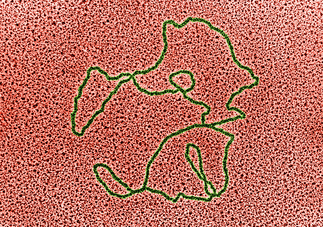
| Color enhanced transmission electron micrograph of a mitochondrial DNA molecule from an oocyte of Xenopus laevis. When disrupted mitochondria are spread by surface tension on a film of water and the liberated DNA is collected on an electron microscope grid,it appears in the form of circular filaments such as that shown here. (Enhancement of 9N1951) | |
| Lizenzart: | Lizenzpflichtig |
| Credit: | Science Photo Library / Fawcett, Don W. |
| Bildgröße: | 4302 px × 3022 px |
| Modell-Rechte: | nicht erforderlich |
| Eigentums-Rechte: | nicht erforderlich |
| Restrictions: |
|
Preise für dieses Bild ab 15 €
Universitäten & Organisationen
(Informationsmaterial Digital, Informationsmaterial Print, Lehrmaterial Digital etc.)
ab 15 €
Redaktionell
(Bücher, Bücher: Sach- und Fachliteratur, Digitale Medien (redaktionell) etc.)
ab 30 €
Werbung
(Anzeigen, Aussenwerbung, Digitale Medien, Fernsehwerbung, Karten, Werbemittel, Zeitschriften etc.)
ab 55 €
Handelsprodukte
(bedruckte Textilie, Kalender, Postkarte, Grußkarte, Verpackung etc.)
ab 75 €
Pauschalpreise
Rechtepakete für die unbeschränkte Bildnutzung in Print oder Online
ab 495 €