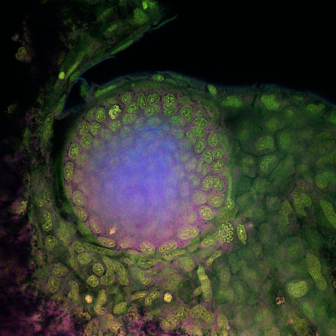
Developing eye
Bildnummer 11875512

| Developing eye of a zebrafish (Danio rerio) embryo,confocal light micrograph. The tissues have been stained with fluorescent dyes and illuminated with a laser. Individual cells (yellow) can be seen dividing. The lens is stained blue (centre). The zebrafish embryo is widely studied by developmental biologists because it is transparent. A confocal microscope detects light only from the focal point of its objective lens. By moving the focal point,sharp images of thin sections of an intact specimen can be obtained | |
| Lizenzart: | Lizenzpflichtig |
| Credit: | Science Photo Library / Reichelt, Stefanie |
| Bildgröße: | 1024 px × 1024 px |
| Modell-Rechte: | nicht erforderlich |
| Eigentums-Rechte: | nicht erforderlich |
| Restrictions: | - |
Preise für dieses Bild ab 15 €
Universitäten & Organisationen
(Informationsmaterial Digital, Informationsmaterial Print, Lehrmaterial Digital etc.)
ab 15 €
Redaktionell
(Bücher, Bücher: Sach- und Fachliteratur, Digitale Medien (redaktionell) etc.)
ab 30 €
Werbung
(Anzeigen, Aussenwerbung, Digitale Medien, Fernsehwerbung, Karten, Werbemittel, Zeitschriften etc.)
ab 55 €
Handelsprodukte
(bedruckte Textilie, Kalender, Postkarte, Grußkarte, Verpackung etc.)
ab 75 €
Pauschalpreise
Rechtepakete für die unbeschränkte Bildnutzung in Print oder Online
ab 495 €
