Coloured SEM of a 6-8 cell segmenting human embryo
Bildnummer 11875045
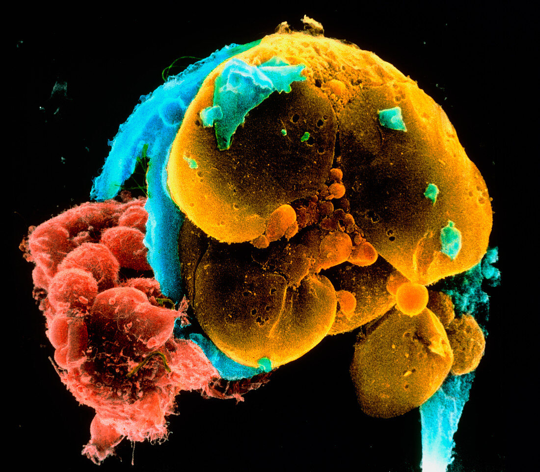
| 6-8 cell embryo. Coloured scanning electron micrograph of layers of a 6-8 cell embryo. The blastomeres (yellow) are the cells formed from divisions of the fertilized egg. Much of the membranous envelope,the zona pellucida (blue),which in life would surround the embryo has been destroyed in preparation. Only fragments of this layer remain. At left a group of the metabolically active cumulus cells has been preserved (red). This cell layer must be penetrated by the sperm in order to fertilize the egg (oocyte). Sperm tails can just be seen at centre left (thin,green). Magnification: x1,500 at 6x7cm size | |
| Lizenzart: | Lizenzpflichtig |
| Credit: | Science Photo Library / PROFESSORS P.M. MOTTA & S. MAKABE |
| Bildgröße: | 4602 px × 4016 px |
| Modell-Rechte: | nicht erforderlich |
| Eigentums-Rechte: | nicht erforderlich |
| Restrictions: | - |
Preise für dieses Bild ab 15 €
Universitäten & Organisationen
(Informationsmaterial Digital, Informationsmaterial Print, Lehrmaterial Digital etc.)
ab 15 €
Redaktionell
(Bücher, Bücher: Sach- und Fachliteratur, Digitale Medien (redaktionell) etc.)
ab 30 €
Werbung
(Anzeigen, Aussenwerbung, Digitale Medien, Fernsehwerbung, Karten, Werbemittel, Zeitschriften etc.)
ab 55 €
Handelsprodukte
(bedruckte Textilie, Kalender, Postkarte, Grußkarte, Verpackung etc.)
ab 75 €
Pauschalpreise
Rechtepakete für die unbeschränkte Bildnutzung in Print oder Online
ab 495 €