Ultrasound of 21 week old foetus's hand with bones
Bildnummer 11875004
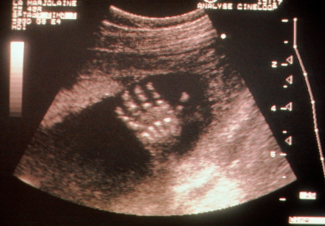
| Hand of a foetus. Ultrasound scan showing the open hand of a 21 week old foetus. Fingers and thumb are each separate and well differentiated,with developing bone (white) seen in the digits and palm. Bone development and ossification starts at 8 weeks of foetal age. This ultrasound scan was made by Dr Philippe Saada. Saada has refined the technique of ultrasound to obtain high-definition images. Ultrasound scanning uses high-frequency sound waves to examine soft tissues such as a foetus inside the womb or to diagnose organ disease. Ultrasound can show whether foetal development is normal. Photographed at the Ultra- sound Centre of La Marjolaine at Montrouge,Paris | |
| Lizenzart: | Lizenzpflichtig |
| Credit: | Science Photo Library / Saada, P. / EURELIOS |
| Bildgröße: | 5072 px × 3520 px |
| Modell-Rechte: | nicht erforderlich |
| Eigentums-Rechte: | nicht erforderlich |
| Restrictions: |
|
Preise für dieses Bild ab 15 €
Universitäten & Organisationen
(Informationsmaterial Digital, Informationsmaterial Print, Lehrmaterial Digital etc.)
ab 15 €
Redaktionell
(Bücher, Bücher: Sach- und Fachliteratur, Digitale Medien (redaktionell) etc.)
ab 30 €
Werbung
(Anzeigen, Aussenwerbung, Digitale Medien, Fernsehwerbung, Karten, Werbemittel, Zeitschriften etc.)
ab 55 €
Handelsprodukte
(bedruckte Textilie, Kalender, Postkarte, Grußkarte, Verpackung etc.)
ab 75 €
Pauschalpreise
Rechtepakete für die unbeschränkte Bildnutzung in Print oder Online
ab 495 €