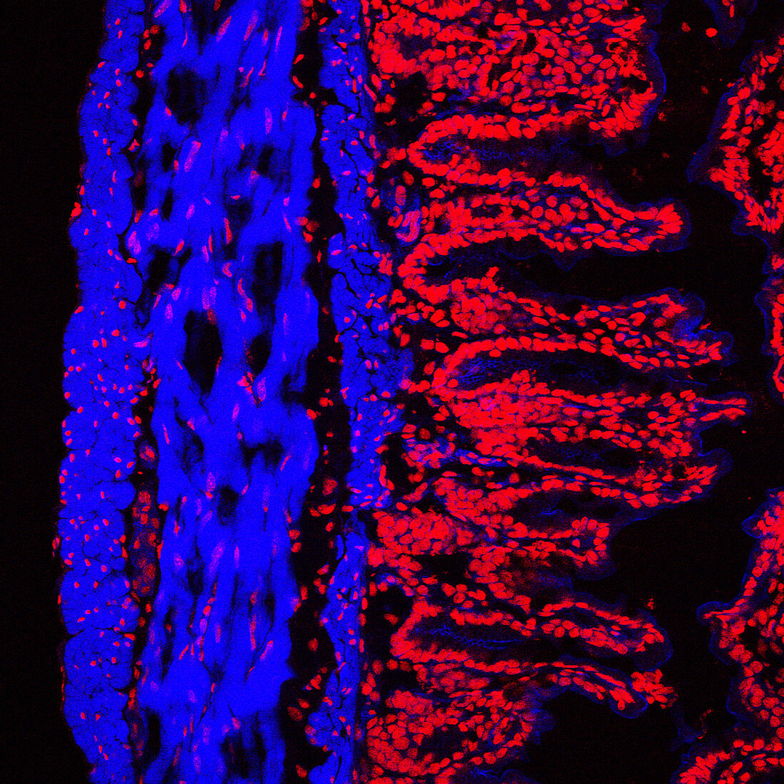
Colon lining
Bildnummer 11873363

| Colon lining. Fluorescence confocal light micrograph of the lining of a mouse colon (large intestine). The colon starts at the small intestine and ends at the rectum. The lumen (interior) of the intestine is at right. The surface (red) of the colon is highly folded,which provides a larger surface area for the absorption of water,the colon's main function. Within the folds are absorptive cells and mucus secreting cells,known as goblet cells. The mucus helps to lubricate the passage of digested food along the colon. Immediately to the left of the folds is a layer of muscle (blue),which contracts to push food along the colon. To the left of that is the submucosal layer (also blue),which consists of fibrous connective tissue and blood vessels | |
| Lizenzart: | Lizenzpflichtig |
| Credit: | Science Photo Library / Schuller, Stephanie |
| Bildgröße: | 2955 px × 2955 px |
| Modell-Rechte: | nicht erforderlich |
| Eigentums-Rechte: | nicht erforderlich |
| Restrictions: | - |
Preise für dieses Bild ab 15 €
Universitäten & Organisationen
(Informationsmaterial Digital, Informationsmaterial Print, Lehrmaterial Digital etc.)
ab 15 €
Redaktionell
(Bücher, Bücher: Sach- und Fachliteratur, Digitale Medien (redaktionell) etc.)
ab 30 €
Werbung
(Anzeigen, Aussenwerbung, Digitale Medien, Fernsehwerbung, Karten, Werbemittel, Zeitschriften etc.)
ab 55 €
Handelsprodukte
(bedruckte Textilie, Kalender, Postkarte, Grußkarte, Verpackung etc.)
ab 75 €
Pauschalpreise
Rechtepakete für die unbeschränkte Bildnutzung in Print oder Online
ab 495 €
Keywords
- Absorption,
- Anatomie,
- anatomisch,
- Biologie,
- biologisch,
- Dickdarm,
- Drüse,
- Drüsen,
- Epithel,
- Falten,
- Fluoreszenz,
- Fluoreszenzlichtmikroskopische Aufnahme,
- fluoreszierend,
- Gefaltet,
- konfokales Lichtmikroskop,
- Lichtmikroskop,
- lichtmikroskopische Aufnahme,
- Maus,
- Muskel,
- Schleim,
- Schleimhaut,
- sekretorisch,
- Tierkörper,
- Verdauungssystem,
- Zelle,
- Zellen
