Tooth enamel formation,SEM
Bildnummer 11872421
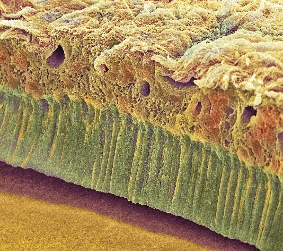
| Tooth enamel formation. Coloured scanning electron micrograph (SEM) of a freeze-fractured section through a tooth,showing the enamel-forming cell layer (green). This epithelium comprises a single layer of columnar cells called ameloblasts. The fracture plane passes up into the tooth from the enamel surface (orange,bottom left). The ameloblast layer has detached from the enamel in which it is normally embedded. Enamel is a hard ceramic layer that covers and protects the teeth. The other end of the ameloblasts originate in the internal tooth tissue (brown,across top). Magnification unknown | |
| Lizenzart: | Lizenzpflichtig |
| Credit: | Science Photo Library / Gschmeissner, Steve |
| Bildgröße: | 3500 px × 3098 px |
| Modell-Rechte: | nicht erforderlich |
| Eigentums-Rechte: | nicht erforderlich |
| Restrictions: | - |
Preise für dieses Bild ab 15 €
Universitäten & Organisationen
(Informationsmaterial Digital, Informationsmaterial Print, Lehrmaterial Digital etc.)
ab 15 €
Redaktionell
(Bücher, Bücher: Sach- und Fachliteratur, Digitale Medien (redaktionell) etc.)
ab 30 €
Werbung
(Anzeigen, Aussenwerbung, Digitale Medien, Fernsehwerbung, Karten, Werbemittel, Zeitschriften etc.)
ab 55 €
Handelsprodukte
(bedruckte Textilie, Kalender, Postkarte, Grußkarte, Verpackung etc.)
ab 75 €
Pauschalpreise
Rechtepakete für die unbeschränkte Bildnutzung in Print oder Online
ab 495 €