Computer image of MEG brain signal plotted on axes
Bildnummer 11870903
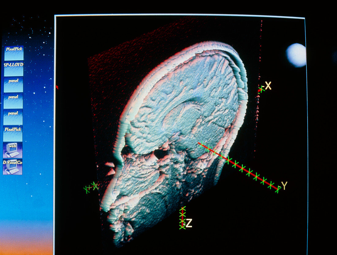
| MEG brain signal. Computer image of a section through the human head (sagittal section),showing a region in the brain at which electrical stimulation has occurred. The region is demarcated in three dimensions with axes X,Y and Z. These axes are a method of representing direction and intensity of an electrical impulse in the brain in response to a stimulus. The impulse was detected using magnetoencephalography (MEG),a brain scanning technique that measures magnetic fields generated from nerve cell activity in the brain. The image of the head was contructed from 3-D MRI scan data. Photographed at New York University Medical Centre in New York City,USA | |
| Lizenzart: | Lizenzpflichtig |
| Credit: | Science Photo Library / Morgan, Hank |
| Bildgröße: | 3632 px × 2750 px |
| Modell-Rechte: | nicht erforderlich |
| Eigentums-Rechte: | nicht erforderlich |
| Restrictions: |
|
Preise für dieses Bild ab 15 €
Universitäten & Organisationen
(Informationsmaterial Digital, Informationsmaterial Print, Lehrmaterial Digital etc.)
ab 15 €
Redaktionell
(Bücher, Bücher: Sach- und Fachliteratur, Digitale Medien (redaktionell) etc.)
ab 30 €
Werbung
(Anzeigen, Aussenwerbung, Digitale Medien, Fernsehwerbung, Karten, Werbemittel, Zeitschriften etc.)
ab 55 €
Handelsprodukte
(bedruckte Textilie, Kalender, Postkarte, Grußkarte, Verpackung etc.)
ab 75 €
Pauschalpreise
Rechtepakete für die unbeschränkte Bildnutzung in Print oder Online
ab 495 €