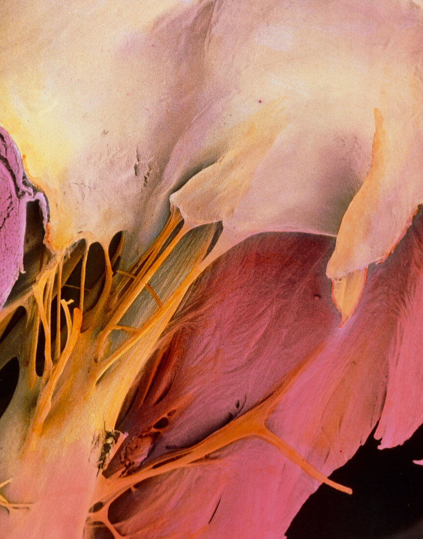
Coloured SEM of the mitral valve of a human heart
Bildnummer 11869001

| Mitral valve of heart. Coloured Scanning Electron Micrograph (SEM) of the mitral valve of a healthy human heart. The mitral valve is one of the atrio- ventricular valves which channels the flow of blood from the left atrium into the left ventricle and prevents any backflow. At top (white) is the mitral valve attached by tendonous cords (chordae tendineae) to a papillary muscle at lower left. Papillary muscles open and close the valve,while the valve's curved surface enables a one way flow of blood. Pink tissue at lower frame is muscle of the ventricle. At top right (pink) is the heart surface. Magnification: x130 at 6x7cm size. Magnification: x170 at 4x5 inch size | |
| Lizenzart: | Lizenzpflichtig |
| Credit: | Science Photo Library / PROFESSORS P.M. MOTTA & G. MACCHIARELLI |
| Bildgröße: | 3828 px × 4875 px |
| Modell-Rechte: | nicht erforderlich |
| Eigentums-Rechte: | nicht erforderlich |
| Restrictions: | - |
Preise für dieses Bild ab 15 €
Universitäten & Organisationen
(Informationsmaterial Digital, Informationsmaterial Print, Lehrmaterial Digital etc.)
ab 15 €
Redaktionell
(Bücher, Bücher: Sach- und Fachliteratur, Digitale Medien (redaktionell) etc.)
ab 30 €
Werbung
(Anzeigen, Aussenwerbung, Digitale Medien, Fernsehwerbung, Karten, Werbemittel, Zeitschriften etc.)
ab 55 €
Handelsprodukte
(bedruckte Textilie, Kalender, Postkarte, Grußkarte, Verpackung etc.)
ab 75 €
Pauschalpreise
Rechtepakete für die unbeschränkte Bildnutzung in Print oder Online
ab 495 €
